• Chemistry Department

NMR Facility - Chemistry Department

Experiment guides, bruker topspin manuals.

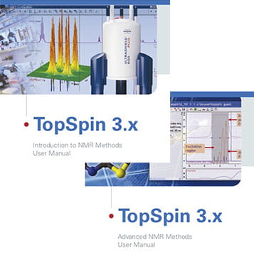
Bruker’s Topspin 3.X manuals offer highly useful step-by-step guides to setting up experiments. If you want to try something new, please feel free to explore these manuals and follow the instructions.

Basic: Introduction to 1D and 2D NMR Methods

Next Step: Advanced NMR Methods

If you have any questions or would like additional orientation, please contact the facility manager .
  • Apply to UW
  • Programs & Majors
  • Cost & Financial Aid
  • Current Students
  • UW Libraries
  • Online Degrees & Programs
  • Degree Plans & Courses
  • Advising & Career Services
  • UW College of Law
  • Honors College
  • Academic Affairs
  • Geological Museum
  • All Colleges
  • Campus Recreation
  • Campus Maps
  • Housing & Dining
  • Transit & Parking
  • University Store
  • Student Organizations
  • Campus Activities
  • Campus Safety
  • Research & Economic Dev.
  • Wyoming INBRE
  • Neuroscience Center
  • Research at AMK Ranch
  • Technology Transfer Office
  • Supercomputing
  • Water Research
  • WY EPSCoR/IDeA
  • American Heritage Center
  • Where We Shine
  • About Laramie
  • Student Stories
  • Campus Fact Book
  • UWYO Magazine
  • Marketing & Brand Center
  • Administrative Resources
  • Strategic Plan
  • +Application Login
  • UW Homepage
  • Instrumentation
  • NMR Facility

NMR Basic Operation - Bruker NMR Spectrometer

  • NMR Service
  • Guidelines for NMR users
  • NMR Basic Operation - Bruker
  • NMR Useful Tips
  • NMR Basic Operation - Anasazi
  • Bruker 300 MHz NMR instrument
  • Bruker 400 MHz NMR instrument
  • Bruker 600 MHz NMR instrument
  • Anasazi EFT-60 NMR instruments
  • Pure Shift NMR
  • Contact and Publications

Chemistry Department

Department 3838

1000 E. University Avenue

Physical Science Bldg 204

Laramie, WY 82071

Phone: 307-766-4363

Fax: 307-766-2807

Email: [email protected]

Find us on Instagram (Link opens a new window)

PDF of Basic Operation

Safety in the NMR laboratory

There are federal training requirements about the dangers of the NMR magnets and the danger of helium asphyxiation that need to be taught to anybody entering the NMR room PS13.

Unauthorized persons need to be supervised at all times by a trained individual who need to instruct them about the dangers. Supervisors assume responsibility for encouraging untrained individuals to circumvent these mandatory requirements.

Please be aware that the superconducting magnets produce very strong magnetic fields that can strongly attract metals and can erase credit cards or other magnetic media. Superconducting magnets are always on. Do not approach the magnets with anything that can influence the magnetic field such as iron keys, tools, wallets, trolleys. People with metallic implants such as heart pacemakers should not approach the magnet without extensive additional safety training. The magnetic field may influence the proper operation of the pacemaker. The magnets in the facility are shielded in such a way that the magnetic field drops off to less than 1 Gauss (0.1 mT) in 2 m. That is considered pretty safe as the earth magnetic field ranges from 0.25 to 0.65 Gauss at the surface. If the magnetic field of the superconducting magnet is disturbed too much, the magnet may suddenly leave the superconducting state and becomes a normal conductor. If such a quench happens, helium evaporates and quickly fills the top of the room, the replacement of oxygen may lead to suffocation. Although the NMR laboratory has been designed in such a way that even the simultaneous quench of all superconducting magnets in the room will leave a layer of breathable air in the lower part of the room, it is strongly recommended to leave the NMR room as soon as possible and to notify the manager. While the temperatures of liquid helium is 4 K (-269 °C), the quench gas temperatures are around 70 - 90 K (600 MHz NMR). Helium gas spreads quickly over the whole room area under the ceiling, and then diffuses away. Due to the low heat capacity of the helium gas, the overall room temperature drop will be barely noticeable. To freeze a pipe, a distance less than 2 - 3 m is needed, less if any media flows in the pipes, or the helium gas does not blow directly on them. Only items directly installed above the magnet or in the path of the quench gas are susceptible to freezing.

Never put any object into the magnet except the NMR tube and spinner. No magnetic stirrer.

Solution Sample Preparation

Please prepare your samples outside the NMR facilities if possible. If you must use the NMR facility, please prepare the samples inside the hood and not on top of the NMR console.

Generally, for 1H-NMR 0.1 mg of sample is sufficent although < 1 mg is usually enough (0.001 mmol /  5 mM) ; for 13C-NMR, 10 mg is sufficient although 0.015 mg is often enough (30 nmol / 0.05 mM). To perform solution-state NMR, the samples are prepared in an appropriate NMR tube and about 0.5-0.6 ml of dissolved sample solution is usually enough. The NMR tubes are commonly 5 mm tubes or 1.7 mm tubes and are available from the Chemistry stockroom. Normally you want to use deuterated solvents such as CDCl 3 or D 2 O to simplify the spectra and to obtain a better signal to noise ratio as protonated solvents show up prominently on proton spectra and may mask the presence of your compound of interest. Also, deuterated solvents can be used to lock the sample (see below) and be useful as an internal reference (see a solvent list here ). If the primary objective is not to observe the protons, but 31P for example, then 5-10% of deuterated solvent is enough (to obtain a lock). Use about 650 ul for common 5 mm NMR tubes (about 51 mm sample height. If it is impractical to use that much, special Shigemi tubes are available that can be filled to 200 ul. Or use 1.7 mm tubes for even less volume (35 ul). It is usually better to not under-fill NMR tubes as NMR setup (shimming) takes much longer and the signal to noise ratio is usually much worse. But over-filling NMR tubes is not a good idea either as it won't improve the signal to noise ratio and consumes laboratory resources needlessly. The sample won't be consumed by the NMR experiment and can be used for other purposes later. The solution should be clear of floating material and well-mixed. If the NMR data shows lines that are broad and/or that have unusual shapes, the main reasons are 1. Insufficient quantity of sample; 2. Significant concentration gradient across sample (inadequate mixing); 3. Floating material (use less concentration or use a better solvent); 4. Presence of paramagnetic compounds (e.g., iron, try to minimize those if possible); 5. Inadequate shimming (don't skip step 8 below).

To clean NMR tubes after the NMR experiments are over, rinse them with water or an organic solvent. Do not use a brush or another abrasive materials. To speed up the drying process, it may be a good idea to use a volatile solvent such as ethanol in the final washing cycle and the NMR tube can be air-dried. It is not recommended to bake the tubes in an oven at more than 45 Degree Celsius as the tubes may deform. A vacuum oven is better. If it is not practical to wait until the NMR tubes are completely dry, use a deuterated solvent such as D 2 O in the final washing cycle. Sample degradation or precipitation may cause material to adhere to the inner walls of the tube. In that case, strong acids such as nitric acid (soaking for 1-3 days) may be employed. Chromic acid is not recommended as the residual chromium often adversely affect NMR experiments. If chromic acid is used nevertheless, nitric acid may remove the residual chromium. For samples that are not dissolved by acid (e.g., some polymers), a solvent that swells the sample may be used and a pipe cleaner might to sufficient to remove the softened material. Agitation in an ultrasonic bath with an appropriate solvent might be useful.

Measuring your sample with the NMR spectrometers

1-dimensional nmr.

Log-on into the NMR computer. If you do not have an account, please contact Dr. Alexander Goroncy to get one. Type topspin<enter> into a terminal program or click the topspin-icon. Please do not log-on to the NMR computer remotely to run topspin as this prevents the user to operate the NMR instrument on-site due to licensing issues!

Before you come anywhere near the magnet, please be sure that you don't carry anything magnetic such as keys, credit cards, screw drivers, pace makers or metallic implants! The magnets may look innocent but they are very powerful indeed and always on!

1. Remove the black cap from the top of the NMR spectrometer.

2. ej<enter> to turn on the eject air flow. Wait until you hear the air flow before proceeding to the next step.

3. Place your NMR tube with spinner in the air flow. Use an appropriate spinner (blue one for most experiments, ceramic one for specialized experiments such as variable temperature experiments). When inserting the NMR tube into the spinner, grap the tube close to the spinner. This will avoid applying a torque that can easily break a tube and drive into a finger. Use the gauge to find the correct sample height in the spinner.Place sample tube in the spinner and the spinner in the same depth gauge. Push or pull the sample tube so that the depth of the sample above and below the center line of the sample depth gauge is equal. However, never exceed the lower limit (position of the adjustable white platform which should be at the line marked 3 mm - 10 mm; do not adjust the height of this white platform). Wipe the tube with tissue such as Kimwipe and after that grap the NMR/spinner combination at the top.

4. ij<enter> to stop the air flow and thereby lower the NMR tube inside the NMR magnet. Wait until it arrives there; wait until topspin says: finished.

5. Open one of your previous experiments in topspin or you can skip this step. It is usually recommended that the first experiment that you do for each new sample is a simple proton experiment such as zg30. There is a long list of possible pulse programs, either in the /opt/topspin3.x./exp/stan/nmr/par folder or in its subfolders. x can be 0, 2, or 5. A list of recommended pulse programs is below.

6. edc<enter> to create a new file. Choose the appropriate pulseprogram (zg30 is for a simple proton experiment) if you want to change it.

7. You may always change the pulse program parameters by typing "rpar" and choosing the desired program. In that case, also type: getprosol<enter>. This loads the default parameters for the particular probe and instrument. The parameters and the pulse program can be inspected under the "AcquPars" tab.

8. rsh<enter> and read a previous shim-file (if you don't know which one, use a recent one with the ending AG).

9. lockdisp<enter> to see the lock display if you don't see it already.

10. lock<enter> and choose solvent from pop-up window. Watch the lock display. When the sample is unlocked, the red/green line will appear at the bottom of the lockdisplay window. When the spectrometer is locking, an FID will appear when the locking finds the correct field setting, and it it will gradually increase to find the correct lock gain settings. The red/green line should be stable in the upper half of the lockdisplay window. You can adjust the position of the line by increasing or decrease the lock gain on the BSMS window (type bsmsdisp<enter>).

11. atma<enter> to tune probe. Also can do it manually with atmm<enter> and use the controls until you get the dip as low as possible (good matching) and directly on the vertical line (good tuning). Manual tuning is recommended for anything other than 1 H and 13 C. Manually tuning usually gives the best results in terms of sensitivity and lineshape but for routine samples the difference is slight. For non-automated tuning probes (like for our 300 MHz NMR spectrometer), the command to use is: wobb<enter>. The capacitors at the probe under the magnet need to be adjusted manually. The NMR probehead must be tuned and matched because it is a resonance circuit. If its resonance frequency and impedance are the same as the transmitter frequency and impedance, respectively, the full transmitter power is transferred to the probehead. However, if either or both are different, part of the transmitter power is reflected by the probehead. This results in a longer 90 Degree pulse. A multi-nuclear probehead has a resonance circuit for each nucleus and each nucleus must be tuned and matched separately. This needs to be done for each sample. Unless the physical conditions of the sample are changing (e.g., chemical reaction, decomposition, temperature), this has to be done only once for each sample.

12. It is generally not recommended to spin the samples. If you do wish to spin it, type "ro on"<enter> or to turn spinning off, "ro off: <enter>. Please be aware that spinning side bands may appear. For multi-dimensional NMR experiment, additional complications appear with spinning samples.

13. topshim<enter> will start the autoshimming routine. For more options: topshim gui<enter>. When Topshim is finished, there will be a message. That may take 2 minutes. For manual shimming: bsmsdisp<enter> and press: shim and adjust the shims manually by observing the red/green line on the lockdisplay. This line generally needs to be as high as possible. If the red/green line reaches the top, it might be necessary to bring it down by reducing the LOCK GAIN. The purpose of shimming is to maximize the magnetic field homogeneity, which depends on probehead and sample geometry. In general, it is necessary to shim the magnetic field after each sample change.

14. getprosol<enter> if you have loaded a default programs above and haven't copied the parameters from an old data set. Prosol is a feature that allows the software to communicate with the probe to determine which probe is installed and to use the standard power and pulse durations for that specific probe.

15. Check parameters such as  ns<enter>, td<enter>, o1p<enter>, sw<enter> for number of scans, number of acquired points, center of the spectrum in ppm, width of the spectrum in ppm. Press the "AcquPars" tab for more settings and press the "Show pulse program parameters" for still more options (second bottom on top left). Both ns, ds should be a multiple of the longest phase cycle in the pulse program to avoid phasing errors after processing. If you are unsure about what that means, either look in the pulseprogram tab and read the description, or at least use multiples of 2, or 4, or 8, or 16, or 32, or 64. For simple 1H-NMR, NS=8 is usually sufficient, and for 13C-NMR, NS=128, for small concentrated chemicals.

16. pulsecal<enter> to calibrate the 90 Degree pulse length. This can be considered optional for a simple proton spectrum, but is important if you wish to do anything more complicated than that. When the pulsecal-routine is finished, it displays the result of the 90 Degree pulse length calibration. If the number is much larger than about 20 ms, then either some of the above steps have been omitted, or, the sample is not of good quality. Often, a substantial amount of undissolved material is the problem.

17. rga<enter> performs auto receiver gain. An abnormal result (e.g., too low, gain=0 will result in no signal) may be due to incorrect parameters settings. Type rg<enter> and type in a lower number in the field such as 2, 4, 8, 16, 32, 64, 128, 256, 512. This is especially important for multi-dimensional experiments to avoid overloading the detector.

18. zg<enter> starts the experiments.

19. Wait until the experiment is finished, or, if you cannot wait that long, type tr<enter> to transfer preliminary data during acquisition. You may type: tr xxx <enter> (such as tr 1024). In that case, the data will be transferred after scan number xxx (such as 1024).

20. efp<enter> and apk<enter>  to process the data. More options can be found in "ProcPars" and "Analyse" in the TopSpin menu bar and should be used if for some reason the automated phase and baseline correction is not working properly. In that case go to "Analyse" and click "Phase", click on the "0" for first order correction and keep it pressed while moving the mouse up and down. Repeat for "1", the first order correction. Don't forget to click "Save".Zooming in can be done with the mouse and the left mouse bottom. To perform an exact zoom via a dialog, type: .zx <enter> and enter the coordinates of the desired region in the dialog box.

For peak integration, the "Integrate" button can be found under "Process". Set the cursor line, starting at the left of the spectrum, to the left of the first peak to be integrated, click the left mouse button and drag the cursor line to the right of the peak, then release the mouse button. Repeat this process to integrate all peaks of interest. It is possible to normalize the integration area. The "Save" button saves the analysis. It is possible to export the spectrum as a PDF or image file by clicking the hard drive button (top left) and choosing

The FID can be manipulated in various ways for sensitivity enhancement or resolution enhancement. This also runs under apodization and window function. Some suggestions how to get started with this are described below. Please type the commands in the command line in topspin.

For sensitivity enhancement : Apply line broadening, enhances first part of FID

lb 2                (positive line broadening, the more positive, the more sensitivity enhancement)

em                (exponential weighted FID)

ft                 (Fourier transform)

apk               (automatic phase correction)

For resolution enhancement : Use Gaussian or sine bell function, changes the shape of the lines of the spectrum, make them narrower, enhances later part of the FID (if used with negative line broadening)

lb -1              (negative line broadening, the more negative, the more resolution enhancement)

gm 0.4            (0<gb<1, the larger gb, the more resolution enhancement)

gm               (Gaussian weighted FID)

"Export". It is also possible to print the spectrum by clicking the hard drive button and "Print".

Most data, including spectrum, FID, peaks, etc., but not parameters can be exported to other applications by selecting the appropriate tab and selecting Edit/Copy from the Topspin menu, the Edit/Paste into the other application.

To export the data (numbers) into another applications, there are several methods. One method is to type "convbin2asc" and the result will be written to the same directory as the original spectrum, e.g. /opt/topspin3.0/data/<user_name>/<sample_name>/<experiment_number>/pdata/<processing_number>/ascii-spec.txt (an example location might be: /home/topspin3.0/data/student/sample/23/pdata/1/ascii-spec.txt ).

Another method to export the data (numbers) is to process the data, then right-click -> Save Display Region to -> A text file for use with other programs -> Please specify destination and type the desired location of your choice. A short way to accomplish this is to simply enter the command "totxt" in the Topspin command line. This will take you directly to the "Please specificy destination" window. This file contains a header with information and only the y values (intensities) are shown and the x values must be generated separetely using the information in the header. This is in contrast with the file that is generated using the "convbin2asc" command that has both x- and y- values generated.

21. halt<enter> will stop the running experiment. This will only be necessary if you wish to prematurely end the experiment. Data is not saved automatically. In case the experiment is run in a repeated loop configuration, the data is saved until the end of the last loop.

22. If you wish to do another experiment with your sample, go back to step 5. You can skip tuning and shimming unless you change the conditions of the sample (e.g., different temperature).

23. If you are finished with your sample, type ej<enter>, take out your sample, type ij<enter>, replace the black cap on top of the spectrometer.

24. Exit topspin and log-out of the computer.

Some pulse programs for routine NMR experiments:

1D proton: zg30, PROTON

1D carbon: zgpg30, C13CPD

1D DEPT-135: dept135, C13DEPT135

1D DEPT-90: dept90, C13DEPT90

You may need to modify the parameters d1 (repetition rate), ns (number of scans), o1p (offset), sw (sweepwidth).

In the Topspin 3 flow interface that is installed in all systems now, there are icons that simply the above steps. In the ACQUIRE tab, there are icons for SAMPLE (insert / eject sample), LOCK (solvent lock), TUNE (tune nuclei), SPIN (turn spin on/off), SHIM (shimming with topshim), PROSOL (getprosol), Gain (automated receiver gain rga), GO (for starting the experiment, zg). For processing, there is a PROCESS icon. If you elect the go this route, please allow sufficient time to finish a job (such as shimming) before going to the next step. Data acquired on the 400 MHz and 600 MHz NMR spectrometers are accessible from the avserver.uwyo.edu computer (in the center of the NMR facility). Please use it to process the data.

2-Dimensional NMR:

All 2D experiments are a series of 1D experiments collected with different timing. They can be divided into two types: homonuclear and heteronuclear. Each type can provide either through-bond (COSY-type) or through space (NOESY-type) decoupling information. They are processed with a Fourier transform in both dimensions.

Experimental setup:

1 Acquire a proton spectrum first and make sure you have recorded the 90 Degree pulse length (step 16 above).

2. Reference the proton spectra and write down the value for the sweep width and center of spectrum. This information is needed for the 2D experiment.

3. If a hetero-nuclear 2D experiment, such as 1H-13C-HSQC or 1H-13C-HMBC is desired, the sweep width for the 13C and the center of the sweep width needs to be set. A 1D 13C-spectrum may be useful to acquire this information.

4. Create a new data set (see step 6 above), and in the "AcquPars" tab load the 2D experiment of your choice from the drop down menu, or copy from a previously acquired dataset.

5. getprosol<enter> if you have loaded a default programs above and haven't copied the parameters from an old data set.

6. Change the parameters for sweep width and offset (o1p and o2p)  in both dimensions with appropriate numbers obtained from the earlier steps. It is always advantageous to reduce the spectra width in both dimensional to the minimum practical value. For the time domain F2, most likely 1H nucleus, you may set 1024, and for the F1 dimension, perhaps 13C, set 128 or 256 if your sample contains many different atoms. Set the 90 Degree 1H pulse width as well. Both ns, ds should be a multiple of the number of phases in the phase program to avoid phasing errors after processing. If you are unsure about what it means, either look in the pulseprogram tab and read the description, or use multiples of 8 or 16.

7. atma<enter> to tune probe. This is only necessary if you haven't tuned the probe with this sample using all the required nuclei before. For example, if you haven't tuned the carbon channel, yet, it is necessary to tune it. For non-automated tuning probes (like for our 300 MHz NMR spectrometer), the command to use is: wobb<enter>. The capacitors at the probe under the magnet need to be adjusted manually.

8. Unless you know what you are doing, don't spin the sample (type "ro off" <enter> if unsure) as otherwise the results may be more complicated to interpret.

9.  rga<enter> performs auto receiver gain. An abnormal result (e.g., too low, gain=0 will result in no signal) may be due to incorrect parameters settings. Type rg<enter> and type in a lower number in the field such as 2, 4, 8, 16, 32, 64, 128, 256, 512. This is especially important for multi-dimensional experiments to avoid overloading the detector. Sometimes the automatic receiver gain optimization does not work very well for 2D experiments as it uses only the first increment to test the receiver gain. Often the signals for subsequent increments are larger and can saturate the receiver. Thus, it is best to use adjust the receiver gain to half or a quarter of the receiver gain that was "optimised" by rga.

10. zg<enter> starts the experiments.

11. Wait until the experiment is finished, or if you cannot wait that long you can get a preview by processing the data and updating the results as new data becomes available.

12. xfb<enter>  to process the data. For more options change the processing parameters in "ProcPars".

13. halt<enter> will stop the running experiment. This will only be necessary if you wish to prematurely end the experiment. Data is saved automatically.

Some useful pulse programs for routine 2-Dimensional NMR experiments:

1H-1H-COSY: cosygpqf

1H-1H-NOESY: noesygppph

1H-13C-HMQC: hmqcgpqf

1H-13C-HSQC: hsqcetgp, hsqcetgpsp, hsqcgpph

1H-13C-HMBC: hmbcgplpndqf

Page created by Alexander Goroncy .

Jump to navigation

Department of Chemistry

  • Discover Chemistry
  • B.A. with major in Chemistry
  • B.S. with major in Chemistry
  • Minor in Chemistry
  • Financial Aid
  • Catalogue Course Listing
  • Course Syllabi Past and Present
  • Research Areas
  • Facilities Overview
  • UVM Chemistry NMR Instrumentation
  • University of Vermont Glassblowing Lab
  • Funded Research Opportunities
  • Outcomes/Careers
  • Chemistry News
  • Upcoming Seminars
  • Department Achievements

College of Arts and Sciences

User guides for bruker nmr spectrometer.

Here are some user guides that will help you with NMR data acquisition on UVM’s Bruker NMR spectrometer.  Please e-mail Monika to request training OR to get help with some of the more complicated experiments.

  • For a basic introduction to acquiring 1H and 13C 1D data see this guide . (Bruker Speedy Setup Guide_v.1.5.pdf)
  • If you’d like to acquire 19F data see this guide . (Bruker-19F1Dacquisition.pdf)
  • And if you’d like to acquire 29Si data see this guide . (Bruker-29Si1Dacquisition.pdf)
  • Determining T1 relaxation values for different 1Hs in your sample is simple.  Please see this guide for doing it on the Bruker NMR (Bruker-T1estimates.pdf).
  • For selective 1D experiments, be it NOESY or TOCSY or a variety of others, see this guide for help (Bruker-Selective1Dexperiments.pdf).
  • If you need to do Variable Temperature (VT) experiments, you should get Monika’s help with this, because depending on whether you want to go hot (easier) or cold (more complicated) there are several steps in the process.  See the Bruker VT guide for this (Bruker-VTexperiments.pdf)
  • Now for solid state NMR experiments, you will need to let Monika know at least a month ahead of time.  The probe gets installed infrequently, liquids users need to be notified and the installation is a fairly complicated process.  This solid state NMR guide will help you with acquiring solids data (Bruker-SolidState-NMR-data-acquisition.pdf)

User guides for Varian NMR spectrometer:

The following user guides will help you with acquiring data on our Varian NMR spectrometer.  Please e-mail Monika to schedule a training or get help.

  • For a basic introduction to acquiring 1H 1D spectra see this guide (Varian-1D-DataAcquisition.pdf).
  • If you’d like to acquire 2D spectra on the Varian, see this guide (Varian-2D-experiment-setup_v1.0.pdf)

Improving the Analysis of Phase-Separated Bio-Fuel Samples with Slice-Selective Total Correlation NMR Spectroscopy

Description.

NMR data supporting "Improving the Analysis of Phase-Separated Bio-Fuel Samples with Slice-Selective Total Correlation NMR Spectroscopy". ABSTRACT: Pyrolysis oil has been identified as a possible alternative fuel source, however widespread use is hindered by high acidity and water content. These negative characteristics can be mitigated by blending with, for example, mixtures of biodiesel, marine gas oil and butanol. These blended biofuel samples can be unstable and often separate into two distinct phases. Analysis of how the components of any blended biofuel samples partition between the two layers is an important step towards understanding the separation process and may provide insight into mitigating the problem. Slice-selective NMR, where the NMR spectrum of only a thin slice of the total sample is acquired, has been previously used to study, non-invasively, bio-oil samples. Here, the technique is extended and improved, with slice-selective two-dimensional correlation experiments used to resolve the distinct chemical spectra of the various components of the phase-separated blended fuel mixtures. EXPERIMENTAL DETAILS: All ¹H NMR measurements were performed on a 300 MHz Bruker Avance spectrometer at 298 K, using a 5 mm BBO probe equipped with a z gradient coil producing a maximum gradient strength of 0.55 T m⁻¹. For the slice-selective NMR experiments, a G4 cascade was used for the selective pulse, with a 5000 Hz bandwidth and applied at offsets of + and − 5000 Hz, corresponding to the upper and lower layers respectively. A gradient of 5 % of the maximum gradient strength was applied concurrently with the selective pulse. This corresponds to a slice 4.3 mm in width, exciting a slice centred 4.3 mm from the centre of the Gz coils. No deuterated solvents were added to the samples. All NMR experiments were acquired without the use of the lock and shimming was achieved using the area of the acquired FID. The data presented here were all acquired with a slice-selective 2D TOCSY experiment with 256 increments, 8 scans and 16 dummy scans, for an experimental duration of 2 hours and 30 minutes. 1D ¹H experiments of all blended samples were also acquired, using 64 scans, for a duration of 16 seconds. All data were processed using TopSpin 2.1. CONTENTS Data organised by samples. Sample A - zg, two slice-selective 1D, 2D TOCSY Sample B - zg, two slice-selective 1D, two slice-selective 2D TOCSY Sample C - zg, two slice-selective 1D, two slice-selective 2D TOCSY Sample D - zg, two (near-identical) slice-selective 1D, 2D TOCSY Sample E - zg, 2D TOCSY Sample F - zg, 2D TOCSY Pulse sequences - 1D data acquired with Bruker_slice_select, 2D TOCSY data acquired with Bruker_slice_select_TOCSY

Steps to reproduce

All data can be processed in TopSpin

Royal Society of Chemistry

Improving the analysis of phase-separated bio-fuel samples with slice-selective total correlation NMR spectroscopy †

ORCID logo

First published on 7th August 2024

Separated samples are a particular challenge for NMR experiments. The boundary is severely detrimental to high-resolution spectra and normal NMR experiments simply add the two spectra of the two layers together. Pyrolysis bio-oils represent an increasingly important alternative fuel resource yet readily separate, whether due to naturally high water content or due to blending, a common practice for producing a more viable fuel. Slice-selective NMR, where the NMR spectrum of only a thin slice of the total sample is acquired, is extended here and improved, with slice-selective two-dimensional correlation experiments used to resolve the distinct chemical spectra of the various components of the phase-separated blended fuel mixtures. Analysis of how the components of any blended biofuel samples partition between the two layers is an important step towards understanding the separation process and may provide insight into mitigating the problem.

Introduction

 
Ω = γG z/2π (1)
 
Δz = (2π/γGB (2)

Only this thin horizontal slice of the sample will be excited by the soft radiofrequency pulse. Before acquisition of the NMR data, the field gradient is switched off and the spectrum of the slice is acquired as in a normal experiment. Slices can be easily moved by changing the offset of the selective pulse. Thinner slices can be acquired, at the expense of reduced signal intensities. The method is not limited to superconducting magnets; so long as the spectrometer has pulsed field gradients in an appropriate geometry, the method described here is transferable.

The use of slice-selective NMR in 1-dimensional chemical applications is a growing field and has been demonstrated in a number of studies, including idealized systems, such as benzene floating on water 2 or water and olive oil mixtures, 3 and the diffusion of small molecules in non-equilibrium systems, such as the mutual diffusion of small molecules, 4 CO 2 in ionic liquids, 5 and small molecules through gels. 6–8 Slice-selective NMR spectroscopy has more recently been utilized in increasingly complex analyses, such as hydrophilic/hydrophobic metabolites, 9 crude oil emulsions, 10 and separated biofuels. 11 Its use is not limited to observing 1 H, with application to the study of 7 Li ions in polymer gels 12,13 and in systems intended to resemble Li-ion batteries. 14

Pyrolysis oils, or bio-oils, are an important example of samples where analysis is often hindered by phase-separation of the samples, yet NMR techniques can give important insight into the nature of the mixture. Pyrolysis is a thermochemical conversion process, involving irreversible heat-driven decomposition of materials, such as lignocellulosic biomass, in the absence of oxygen. 15 The pyrolysis products typically contain char, gases, and an oil. The oil is a potential fuel, but typically cannot be used directly in unmodified engines as it contains too much water and various other oxygen-containing species present 16 render it too acidic. There are several methods by which the utility of a pyrolysis product can be improved, 17–19 such as by blending with other products. 20–22 Such multiple component blends are typically opaque and can readily separate into a multiple-phase solution. 23–25 Once separated, the blends are highly unsuitable as fuel products and could cause significant damage to an engine if used. A key challenge to the successful blending of these fuel products is the analysis, understanding and mitigation of this phase separation. The NMR analysis of pyrolysis oils, also known as bio-oils, is well-established and comprehensive reviews are available. 26,27 However, any analysis of these oils, blended or otherwise, is complicated by the large number of species present and the range of functional groups that may be present.

Here, the improved performance of slice-selective NMR analysis of complex phase-separated samples, such as pyrolysis bio-oils and their blends, is demonstrated by combining slice-selective methods with two-dimensional NMR techniques. Total correlation spectroscopy (TOCSY) is used here, as the final spectra produced can be phased to give pure absorption mode peaks.

Experimental

Methods and materials.

Sample Component composition (%)
Bio-oil Butanol FAME Marine gas oil
A 20 40 40 0
B 30 40 0 30
C 30 20 50 0
D 10 80 10 0
E 20 50 7.5 22.5
F 30 40 15 15

NMR experiments

Results and discussion, 2d 1 h tocsy of un-separated bio-oil blends.

2D H TOCSY spectra of three-component unseparated sample A. Gray inset details peaks along horizontal row at ca. 5.3 ppm.

However, the TOCSY spectrum reveals additional components of the mixture, both expected and unexpected. The fatty acid methyl ester can also be observed, as a row of resonances horizontally or vertically along 5.4 ppm. With the long alkyl chain on the fatty acid methyl ester, a suitably long spin-lock is needed to couple together the most distant protons on the chain. The signals highlighted in the inset confirm that the spin-lock period selected is appropriate for the sample. While the intensity of the methyl peak is low, particularly compared with the more plentiful methylene signals, it does appear along the same horizontal line as the other FAME signals. Additional peaks, belonging to neither butanol nor fatty acid methyl ester are observed as cross peaks between ca. 1.5 ppm and ca. 2 ppm. This spectrum shows the advantages of the TOCSY pulse sequence. The complete NMR spectrum of individual components can be readily resolved.

Two-dimensional TOCSY spectra of an additional unseparated, three-component and two unseparated, four-component, samples, samples D, E and F, are presented in the ESI as Fig. S2–S4. † While these all contain different amounts of bio-oil, butanol, marine gas oil and FAME, their spectra share many of the features of sample A revealed in Fig. 1 .

Slice-selective 2D 1 H TOCSY of separated samples

2D H TOCSY spectra of three-component separated sample B. Left-hand spectrum depicts the upper layer and right-hand spectrum depicts the lower layer. Spectral regions indicated by boxes are reproduced, enlarged, in .

In order to make a more detailed comparison between the two layers, the regions from 3 to 8 ppm, indicated by boxes in Fig. 2 , are magnified and overlaid. This comparison is depicted in Fig. 3 , with the upper layer in blue and the lower layer in red. This presentation of the two-dimensional TOCSY spectra makes the differences between the two layers easily visible. The cross peaks between ca. 7 ppm and ca. 2 ppm in the upper layer spectrum confirms the presence aromatic species with alkyl substituents. The broad nature of these peaks indicates a wide range of species, likely polymeric or fused ring systems. As these species are found in the upper, oil, layer, they are likely to be heavier fractions of the marine gas oil. On the other hand, the cross peaks in the lower layer are both smaller in area and are clustered around 3.5 and 5.5 ppm, indicating a large number of distinct, smaller compounds with both polar functional groups and unsaturated, olefinic, chains. With pyrolysis bio-oil being produced from lignocellulosic biomass, the presence of compounds with structures based on monolignols or the constituent units of lignin is expected.

2D H TOCSY spectrum between 2 and 8 ppm (region indicated by box in ) of three-component separated sample B. Blue spectrum indicates upper layer while red spectrum indicates lower layer. The individual slice-selective 2D TOCSY spectra of each layer are reproduced in ESI as Fig. S5 and S6.

Fig. 4(a) depicts overlaid 2D TOCSY spectra of both the upper layer, in blue, and the lower layer, in red, of a final separated, three component sample (sample C). In this figure, a large range of chemical shifts with a broad dynamic range is depicted and the contour levels of the 2D plots have been adjusted to show as full a range of smaller, less intense, peaks as possible. These peaks are particularly evident in the lower layer, with a large number of small, sharp cross peaks between 3 and 5 ppm. These indicates that, practically, all of the bio-oil components are found in the aqueous layer.

(a) Slice-selective 2D H TOCSY spectra of both upper and lower layers of three-component separated sample C. Blue spectrum indicates upper layer. Red spectrum indicates lower layer. Along the x- and y-axes, the blue and red 1D spectra depict the slice-selective spectra of the upper and lower layers, respectively. A 1D spectrum of the whole sample has been overlaid at the bottom, in black, for further comparison. (b) Slice-selective 2D H TOCSY spectrum of lower layer of three-component separated sample C.

Butanol is again partitioned between the upper and lower layers, more evenly than in the previous example, with ca. 50% in each layer. Identification and measurement of the butanol peaks is made easier by the improved resolution of the spectrum. This sample contains no marine gas oil. These observations are confirmed in Fig. 4(b) , which depicts the 2D TOCSY of only the lower slice of the final separated sample for an expanded chemical shift range.

As with every 2D spectrum of the lower, aqueous, layer, there are many sharp peaks, with numerous cross peaks observed between 3 and 5 ppm. What are likely to be small chain alcohols can be observed the shadow of the intense butanol peaks at ca. 4 ppm. In addition, the horizontal lines along ca. 5.5 ppm suggests the presence of olefinic groups. Each distinct cross peak in the two-dimensional spectrum corresponds to coupling between two distinct proton environments within the same spin system. Further analysis and identification of individual components in the bio-oil component of the mixture could be achieved through use of machine learning tools applied to this large set of NMR data.

Conclusions

This paper demonstrates the improved analysis of phase-separated samples by the successful implementation of slice-selective two-dimensional TOCSY NMR. Blended bio-oil samples are often unstable, separating into two distinct, often opaque, phases. By extending the spectra into a second dimension, the resolution of individual peaks are significantly improved and it is easier to identify specific species in the different layers of the sample. In addition, coupling information is now revealed, allowing for identification of more components in the mixtures and tentative assignments of compounds present. Improved analysis of how the components of any blended biofuel samples partition between the two layers is an important step towards understanding the separation processes and may provide insight into mitigating the problem. With the wider use of NMR techniques in studying biofuel samples as well as other separated or heterogeneous samples, slice-selective NMR techniques offer a powerful, additional analytical tool.

Data availability

Author contributions, conflicts of interest, acknowledgements.

  • J. N. Dumez, Prog. Nucl. Magn. Reson. Spectrosc. , 2018, 109 , 101  CrossRef   CAS   PubMed .
  • J. Lambert, R. Hergenroder, D. Suter and V. Deckert, Angew. Chem., Int. Ed. , 2009, 48 , 6343  CrossRef   CAS   PubMed .
  • C. Mantel, P.-A. Bayle, S. Hediger, C. Berthon and M. Bardet, Magn. Reson. Chem. , 2010, 48 , 600  CrossRef   CAS   PubMed .
  • C. F. Pantoja, J. A. Bolanos, A. Bernal and J. Wist, Magn. Reson. Chem. , 2017, 55 , 519  CrossRef   CAS   PubMed .
  • J. Allen and K. Damodaran, Magn. Reson. Chem. , 2015, 53 , 200  CrossRef   CAS   PubMed .
  • C. Garcia-Aparicio, I. Quijada-Garrido and L. Garrido, J. Colloid Interface Sci. , 2012, 368 , 14  CrossRef   CAS   PubMed .
  • Y. Mitrev, S. Simova and D. Jeannerat, Chem. Commun. , 2016, 52 , 5418  RSC .
  • M. A. Wisniewska and J. G. Seland, J. Colloid Interface Sci. , 2019, 533 , 671  CrossRef   CAS   PubMed .
  • R. Evans, A. Sandhu, A. V. Bridgwater and K. Chong, Energy Fuels , 2017, 31 , 4135  CrossRef   CAS .
  • T. N. Hjartnes, G. H. Sørland, S. Simon and J. Sjöblom, Ind. Eng. Chem. Res. , 2019, 58 , 2310  CrossRef   CAS .
  • K. Seeger, Anal. Chem. , 2021, 93 , 1451  CrossRef   CAS   PubMed .
  • A. C. Poppler, S. Frischkorn, D. Stalke and M. John, ChemPhysChem , 2013, 14 , 3103  CrossRef   PubMed .
  • T. Niklas, D. Stalk and M. John, Chem. Commun. , 2015, 51 , 1275  RSC .
  • S. A. Krachkovskiy, A. D. Pauric, I. C. Halalay and G. R. Goward, J. Phys. Chem. Lett. , 2013, 4 , 3940  CrossRef   CAS .
  • A. V. Bridgwater, Biomass Bioenergy , 2012, 38 , 68  CrossRef   CAS .
  • D. Mohan, C. U. Pittman Jr and P. H. Steele, Energy Fuels , 2006, 20 , 848  CrossRef   CAS .
  • E. Butler, G. Devlin, D. Meier and K. A. McDonnell, Renewable Sustainable Energy Rev. , 2011, 15 , 4171  CrossRef   CAS .
  • P. M. Mortensen, J. D. Grunwaldt, P. A. Jensen, K. G. Knudsen and A. D. Jensen, Appl. Catal., A , 2011, 407 , 1  CrossRef   CAS .
  • A. H. Zacher, M. V. Olarte, D. M. Santosa, D. C. Elliott and S. B. Jones, Green Chem. , 2014, 16 , 491  RSC .
  • M. Garcia-Perez, J. Shen, X. S. Wang and C. Z. Li, Fuel Process. Technol. , 2010, 91 , 296  CrossRef   CAS .
  • T. Y. Kim, S. Lee and K. Kang, Energy , 2015, 93 , 2241  CrossRef   CAS .
  • M. Zhang and H. Wu, Energy Fuels , 2014, 28 , 4650  CrossRef   CAS .
  • A. Krutof and K. Hawboldt, Renewable Sustainable Energy Rev. , 2016, 59 , 406  CrossRef   CAS .
  • K. J. Chong and A. V. Bridgwater, Environ. Prog. Sustainable Energy , 2017, 36 , 677  CrossRef   CAS .
  • A. Alcala and A. V. Bridgwater, Fuel , 2013, 109 , 417  CrossRef   CAS .
  • C. A. Mullen, G. D. Strahan and A. A. Boateng, Energy Fuels , 2009, 23 , 2707  CrossRef   CAS .
  • N. Hao, H. Ben, C. G. Yoo, S. Adhikari and A. J. Ragauskas, Energy Fuels , 2016, 30 , 6863  CrossRef   CAS .
  • C.-Y. Lin, Energies , 2013, 6 , 4945  CrossRef .
  • L. Emsley and G. Bodenhausen, Chem. Phys. Lett. , 1990, 165 , 469  CrossRef   CAS .
  • D. M. Doddrell, D. T. Pegg and M. R. Bendall, J. Magn. Reson. , 1982, 48 , 323  CAS .
  • J. B. Nielsen, A. Jensen, C. B. Schandel, C. Felby and A. D. Jensen, Sustainable Energy Fuels , 2017, 1 , 2006  RSC .
  • D. H. Lysak, W. Bermel, V. Moxley-Paquette, C. Michal, R. Ghosh-Biswas, R. Soong, B. Nashman, A. Lacerda and A. J. Simpson, Anal. Chem. , 2023, 95 , 14392  CrossRef   CAS   PubMed .
  • R. Evans, Z. Deng, A. K. Rogerson, A. S. McLachlan, J. J. Richards, M. Nilsson and G. A. Morris, Angew. Chem., Int. Ed. , 2013, 52 , 3199  CrossRef   CAS   PubMed .
  • W. Li, G. Kagan, R. Hopson and P. G. Williard, J. Chem. Educ. , 2011, 88 , 1331  CrossRef   CAS .
  • N. A. Halliday, A. C. Peet and M. M. Britton, J. Phys. Chem. B , 2010, 114 , 13745  CrossRef   CAS   PubMed .
  • N. Esturau and J. F. Espinosa, J. Org. Chem. , 2006, 71 , 4103  CrossRef   CAS   PubMed .
  • J. A. Aguilar, M. Nilsson, G. Bodenhausen and G. A. Morris, Chem. Commun. , 2012, 48 , 811  RSC .
Electronic supplementary information (ESI) available: Pulse sequences used and additional NMR spectra. See DOI:

1 H, 13 C and 15 N resonance assignments of a shark variable new antigen receptor against hyaluronan synthase

  • Published: 14 August 2024

Cite this article

bruker nmr experiments

  • Yuxin Liu 1 , 2 , 3 ,
  • Hao Wang 2 , 3 ,
  • Cookson K. C. Chiu 4 ,
  • Yujie Wu 4 &
  • Yunchen Bi 2 , 3  

Single domain antibody (sdAb) is only composed of a variable domain of the heavy-chain-only antibody, which is devoid of light chain and naturally occurring in camelids and cartilaginous fishes. Variable New Antigen Receptor (VNAR), a type of single domain antibody present in cartilaginous fishes such as sharks, is the smallest functional antigen-binding fragment found in nature. The unique features, including flexible paratope, high solubility and outstanding stability make VNAR a promising prospect in antibody drug development and structural biology research. However, VNAR’s research has lagged behind camelid-derived sdAb, especially in the field of structural research. Here we report the 1 H, 15 N, 13 C resonance assignments of a VNAR derived from the immune library of Chiloscyllium plagiosum , termed B2-3, which recognizes the hyaluronan synthase. Analysis of the backbone chemical shifts demonstrates that the secondary structure of VNAR is predominately composed of β-sheets corresponding to around 40% of the B2-3 backbone. The Cβ chemical shift values of cysteine residues, combined with mass spectrometry data, clearly shows that B2-3 contains two pairs of disulfide bonds, which is import for protein stability. The assignments will be essential for determining the high resolution solution structure of B2-3 by NMR spectroscopy.

This is a preview of subscription content, log in via an institution to check access.

Access this article

Subscribe and save.

  • Get 10 units per month
  • Download Article/Chapter or eBook
  • 1 Unit = 1 Article or 1 Chapter
  • Cancel anytime

Price includes VAT (Russian Federation)

Instant access to the full article PDF.

Rent this article via DeepDyve

Institutional subscriptions

bruker nmr experiments

Explore related subjects

Data availability.

The assigned 1 H, 15 N and 13 C chemical shift of B2-3 VNAR has been deposited in the BioMagResBank ( http://www.bmrb.wisc.edu/ ) under the accession number 52,417.

Berjanskii MV, Wishart DS (2005) A simple method to predict protein flexibility using secondary Chemical shifts. J Am Chem Soc 127(43):14970–14971. https://doi.org/10.1021/ja054842f

Article   Google Scholar  

Delaglio F, Grzesiek S, Vuister GW, Zhu G, Pfeifer J, Bax A (1995) NMRPipe: a multidimensional spectral processing system based on UNIX pipes. J Biomol NMR 6(3):277–293. https://doi.org/10.1007/BF00197809

Diaz M, Greenberg AS, Flajnik MF (1998) Somatic hypermutation of the new antigen receptor gene (NAR) in the nurse shark does not generate the repertoire: possible role in antigen-driven reactions in the absence of germinal centers. Proc Natl Acad Sci U S A 95(24):14343–14348. https://doi.org/10.1073/pnas.95.24.14343

Article   ADS   Google Scholar  

Greenberg AS, Avila D, Hughes M, Hughes A, McKinney EC, Flajnik MF (1995) A new antigen receptor gene family that undergoes rearrangement and extensive somatic diversification in sharks. Nature 374(6518):168–173. https://doi.org/10.1038/374168a0

Hamers-Casterman C, Atarhouch T, Muyldermans S, Robinson G, Hamers C, Songa EB, Bendahman N, Hamers R (1993) Naturally occurring antibodies devoid of light chains. Nature 363(6428):446–448. https://doi.org/10.1038/363446a0

Liu W, Song H, Chen Q, Yu J, Xian M, Nian R, Feng D (2018) Recent advances in the selection and identification of antigen-specific nanobodies. Mol Immunol 96:37–47. https://doi.org/10.1016/j.molimm.2018.02.012

Markley JL, Bax A, Arata Y, Hilbers CW, Kaptein R, Sykes BD, Wright PE, Wuthrich K (1998) Recommendations for the presentation of NMR structures of proteins and nucleic acids. IUPAC-IUBMB-IUPAB Inter-union Task Group on the Standardization of Data Bases of Protein and nucleic acid structures determined by NMR spectroscopy. J Biomol NMR 12(1):1–23. https://doi.org/10.1023/a:1008290618449

Muyldermans S (2013) Nanobodies: natural single-domain antibodies. Annu Rev Biochem 82:775–797. https://doi.org/10.1146/annurev-biochem-063011-092449

Muyldermans S (2021) Applications of Nanobodies. Annu Rev Anim Biosci 9:401–421. https://doi.org/10.1146/annurev-animal-021419-083831

Pardon E, Laeremans T, Triest S, Rasmussen SG, Wohlkonig A, Ruf A, Muyldermans S, Hol WG, Kobilka BK, Steyaert J (2014) A general protocol for the generation of Nanobodies for structural biology. Nat Protoc 9(3):674–693. https://doi.org/10.1038/nprot.2014.039

Shen Y, Bax A (2013) Protein backbone and sidechain torsion angles predicted from NMR chemical shifts using artificial neural networks. J Biomol NMR 56(3):227–241. https://doi.org/10.1007/s10858-013-9741-y

Ubah OC, Lake EW, Gunaratne GS, Gallant JP, Fernie M, Robertson AJ, Marchant JS, Bold TD, Langlois RA, Matchett WE, Thiede JM, Shi K, Yin L, Moeller NH, Banerjee S, Ferguson L, Kovaleva M, Porter AJ, Aihara H, LeBeau AM, Barelle CJ (2021) Mechanisms of SARS-CoV-2 neutralization by shark variable new antigen receptors elucidated through X-ray crystallography. Nat Commun 12(1):7325. https://doi.org/10.1038/s41467-021-27611-y

Wei L, Wang M, Xiang H, Jiang Y, Gong J, Su D, Al Azad MAR, Dong H, Feng L, Wu J, Chan LL, Yang N, Shi J (2021) Bamboo shark as a small animal model for single domain antibody production. Front Bioeng Biotechnol 9:792111. https://doi.org/10.3389/fbioe.2021.792111

Zielonka S, Empting M, Grzeschik J, Konning D, Barelle CJ, Kolmar H (2015) Structural insights and biomedical potential of IgNAR scaffolds from sharks. MAbs 7(1):15–25. https://doi.org/10.4161/19420862.2015.989032

Download references

Acknowledgements

We thank for grant supports from the National Natural Science Foundation of China (Grant Number: 42376136), Research on Simulation Technology and Device of Key Processes of Typical Marine Ecological Disasters in the Pre-Research Project of Major Scientific Facilities in Shandong Province (DKXZZ202205). We thank the staffs at the Intelligent Simulator of Marine Ecosystems, ISME and the staffs at the mass spectrometry system at the Shenzhen Bay Laboratory for instrument support and technical assistance. This work is also supported by Oceanographic Data Center, IOCAS.

Author information

Authors and affiliations.

College of Life Sciences, Qingdao University, Qingdao, China

CAS and Shandong Province Key Laboratory of Experimental Marine Biology, Institute of Oceanology, Center for Ocean Mega-Science, Chinese Academy of Sciences, Qingdao, China

Yuxin Liu, Hao Wang & Yunchen Bi

Laboratory for Marine Biology and Biotechnology, Qingdao Marine Science and Technology Center, Qingdao, China

Shenzhen Bay Laboratory, Shenzhen, China

Cookson K. C. Chiu & Yujie Wu

You can also search for this author in PubMed   Google Scholar

Contributions

Y.L. , C.C. and Y.W. performed the experiments. H.W. and Y.B. wrote the main manuscript text and H.W. prepared figures. All authors reviewed the manuscript.

Corresponding authors

Correspondence to Yujie Wu or Yunchen Bi .

Ethics declarations

Competing interests.

The authors declare no competing interests.

Additional information

Publisher’s note.

Springer Nature remains neutral with regard to jurisdictional claims in published maps and institutional affiliations.

Electronic supplementary material

Below is the link to the electronic supplementary material.

Supplementary Material 1

Rights and permissions.

Springer Nature or its licensor (e.g. a society or other partner) holds exclusive rights to this article under a publishing agreement with the author(s) or other rightsholder(s); author self-archiving of the accepted manuscript version of this article is solely governed by the terms of such publishing agreement and applicable law.

Reprints and permissions

About this article

Liu, Y., Wang, H., Chiu, C.K.C. et al. 1 H, 13 C and 15 N resonance assignments of a shark variable new antigen receptor against hyaluronan synthase. Biomol NMR Assign (2024). https://doi.org/10.1007/s12104-024-10190-6

Download citation

Received : 17 May 2024

Accepted : 01 August 2024

Published : 14 August 2024

DOI : https://doi.org/10.1007/s12104-024-10190-6

Share this article

Anyone you share the following link with will be able to read this content:

Sorry, a shareable link is not currently available for this article.

Provided by the Springer Nature SharedIt content-sharing initiative

  • Single domain antibody
  • NMR spectroscopy assignments
  • Find a journal
  • Publish with us
  • Track your research

IMAGES

  1. The Bruker NMR equipment used for phosphorus-31 and fluorine-19 MAS NMR

    bruker nmr experiments

  2. NMR Instruments

    bruker nmr experiments

  3. Bruker’s high-performance Avance III NMR spectrometer technology

    bruker nmr experiments

  4. 395 GHz/600 MHz Bruker Avance III DNP NMR

    bruker nmr experiments

  5. AV 400

    bruker nmr experiments

  6. Bruker Advances NMR Magnet Portfolio, Innovates in Industrial Solutions

    bruker nmr experiments

COMMENTS

  1. PDF Basic 1D and 2D Experiments

    experiments and processes the acquired NMR data. 1.3 Bruker NMR software There are three major tasks that are controlled by the NMR software: acquisition, processing and plotting. The TopSpin program is the user interface for all of these tasks. Compared to the former XWIN-NMR software,

  2. NMR Instruments

    Nuclear magnetic resonance spectroscopy (NMR) is an analytical technique that allows for non-invasive and non-destructive plus quantitative analytical investigations into molecular structure, dynamic processes and chemical reactions. Bruker is the industry leader in the manufacturing of unique high-performance magnetic resonance instruments.

  3. PDF 1D and 2D Experiments Step-by-Step Tutorial

    DEPT-135 Experiment User Guide Version 002 BRUKER BIOSPIN 17 (123) Figure 2.14. 6. Click on 'File' and select 'Print' by clicking on it DEPT-135 Experiment 2.4 Sample: 30 mg Brucine in CDCl3 Experiment set up 2.4.1 NOTE: This experiment usually follows a regular 1H decoupled 13C experiment.

  4. PDF PULSE PROGRAM CATALOGUE: I. 1D & 2D NMR EXPERIMENTS

    Pulse Program Catalogue NMRGuide 4.1 - Topspin 2.0 BRUKER Pulse Program Catalogue written by Teodor Parella This catalogue presents the pulse sequence diagram for all standard pulse programs included

  5. Fragment screening by ligand observed nmr

    Conclusion. Ligand observed NMR is an automated fragment screening technique that yields high quality hits in primary and secondary screens. Nowadays, parameter sets with optimized measurement conditions are available which significantly facilitates the setup of NMR-based screening experiments, even for novice users.

  6. Solid-State NMR

    In addition, Bruker offers a wide variety of specialty probes, e.g. for solid-state NMR experiments at extreme temperatures or probes which are intended for battery research. Bruker's extensive probe portfolio is complemented by high-quality consumables and advanced automation solutions. Please contact us to discuss your particular application.

  7. 1D NOESY made easy

    NOESY experiments always include a kinetic aspect. The NOE builds up during a delay in the experiment called the "mixing time". By convention, this delay is D8 in Bruker pulse sequences. It is typically set to around 600 msec for small molecules. Magnetization transfer by NOE between spins can be quantified.

  8. PDF Estimating T1s for Hs in your sample on the Bruker NMR

    on the Bruker NMRby Monika Ivancic, PhD (April, 2020)Estimates of T1 relaxation values of different 1Hs in your sample are important for setting up more advanced NMR experi. nts, and essential when quantitative work is desired. The repetition time in any NMR experiment is determined by the r. laxation. elay (D1) and the acquisition time (AQ ...

  9. Experiments

    Bruker Topspin Manuals. Bruker's Topspin 3.X manuals offer highly useful step-by-step guides to setting up experiments. If you want to try something new, please feel free to explore these manuals and follow the instructions. Basic: Introduction to 1D and 2D NMR Methods. Next Step: Advanced NMR Methods.

  10. PDF Step By Step Instructions for 2D NMR Experiments on the Bruker 300 MHz

    o1p, and SR values for the carbon experiment. (8) Create a new data set using the NMR step by step drop down menu. Again, use the same . name for the experiment as you did in the previous steps but use a different experiment . number (ex. 3 or 4 or 5). (9) Load the 2D experiment of your choice from the 2D experiments list in the drop down menu.

  11. PDF BRUKER NMR TRAINING v. i

    BRUKER NMR TRAINING Magnetic Resonance Research Center of the University of Notre Dame 8/18/2022 This is a beginner user guide for collecting basic proton and carbon 1D NMR experiments on Bruker instruments at MRRC. If you have any questions, please, do not hesitate to email NMR staff at [email protected] Safety

  12. Bruker Tutorials

    Bruker Tutorials. This tutorial will guide users through the process of data collection and analysis for a small molecule using Bruker TopSpin3.0 or newer. Each step will contain an explanation of input and output, using typical settings for general use. Click on the experiment that you are interested in and follow the link:

  13. PDF Selective 1D Experiments on the ruker NMR

    Selective 1D Experiments on the ruker NMR (1D NOESY, 1D TOSY, etc) by Monika Ivancic, PhD (December, 2018) 1. Acquire a 1H 1D experiment (uvm.1H1D) (see ruker Speedy setup guide) DO NOT spin the sample! (OR turn spin off after you acquire the 1H 1D) 2. Determine T1 relaxation times for the 1Hs of interest in your sample (see Estimating 1H T1

  14. Advanced NMR Methods

    Formerly AVANCE II. This 3 day course covers theoretical and experimental details of many aspects of common NMR experiments such as gradients, shaped pulses and solvent suppression. The morning sessions are comprised of classroom lectures followed by hands on sessions at spectrometer. Additional classroom and lab session topics covered during ...

  15. PDF VT experiments on Bruker NMR spectrometer

    Low temperature experiments th (by Monika Ivancic, October 20 , 2016) There is no chiller on the Bruker NMR spectrometer, so if you absolutely must use the Bruker to go cold, you will need to use the small LN2 dewar with the LN2 Exchanger for any temperature below ambient. The Varian NMR does have a chiller, so if you need temps

  16. PDF Kinetic NMR Experiments

    Kinetic NMR Experiments. When doing an NMR kinetics experiment, one wants to take a series of spectra over a period of time. There are two possible ways to do this: with a series of independent 1D spectra or in pseudo 2D mode. The 2D mode is greatly preferred because the timing between spectra is 100% accurate and data can be analyzed with both ...

  17. NMR Basic Operation

    Data acquired on the 400 MHz and 600 MHz NMR spectrometers are accessible from the avserver.uwyo.edu computer (in the center of the NMR facility). Please use it to process the data. 2-Dimensional NMR: All 2D experiments are a series of 1D experiments collected with different timing.

  18. PDF Solvent Suppression using TopSpin 3

    Bruker's LC-NMR experiments use noesy1d-presat, and are implemented in the facility in IconNMR; see this document for more . Solvent Suppression using TopSpin 3.x . ... two) on these 1D experiments. July 2022: see NMR staff for updates that include LC-NMR experiments. A. Initial Setup: 1. Always start by acquiring a one-scan 1

  19. New in Bruker User Library: a Family of Experiments for Fast

    Multidimensional TOCSY and NOESY lie at the foundation of homonuclear NMR correlation experiments. Low sensitivity is an intrinsic downside of these polarization transfer methods -particularly for systems involving fast relaxing or rapidly exchanging, labile protons. We have recently introduced Looped PROjected SpectroscopY.

  20. User Guides for Bruker NMR spectrometer

    Here are some user guides that will help you with NMR data acquisition on UVM's Bruker NMR spectrometer. Please e-mail Monika to request training OR to get help with some of the more complicated experiments. For a basic introduction to acquiring 1H and 13C 1D data see this guide. (Bruker Speedy Setup Guide_v.1.5.pdf)

  21. Improving the Analysis of Phase-Separated Bio-Fuel Samples with Slice

    EXPERIMENTAL DETAILS: All ¹H NMR measurements were performed on a 300 MHz Bruker Avance spectrometer at 298 K, using a 5 mm BBO probe equipped with a z gradient coil producing a maximum gradient strength of 0.55 T m⁻¹. ... All NMR experiments were acquired without the use of the lock and shimming was achieved using the area of the acquired ...

  22. Improving the analysis of phase-separated bio-fuel samples with slice

    NMR experiments All 1 H NMR measurements were performed on a 300 MHz Bruker Avance spectrometer at 298 K, using a 5 mm BBO probe equipped with a z gradient coil producing a maximum gradient strength of 0.55 T m −1.For the slice-selective NMR experiments, a G4 cascade 29 was used for the selective pulse, with a 5000 Hz bandwidth and applied at offsets of + and −5000 Hz, corresponding to the ...

  23. DNP-NMR

    Bruker's DNP NMR spectrometers are available for solid-state NMR corresponding to 1 H frequencies of 400 to 900 MHz. Introduction to DNP-NMR. Dynamic Nuclear Polarization (DNP) is a technique used to enhance the sensitivity of NMR experiments. It involves the transfer of polarization from highly polarized electron spins to the nuclear spins ...

  24. 1H, 13C and 15N resonance assignments of a shark variable ...

    The NMR spectra were acquired at 298 K on Bruker Avance NEO 600 MHz (with a cryoprobe), equipped with four RF channels and a triple-resonance probe with pulsed field gradients. ... The triple resonance NMR experiments led to an assignment of 94.2% % (97 residues) of the observed signals. The missing residues were M1, A2, L33, T46, L51, G55, T63 ...

  25. New in Bruker User Library: Manchester Experiments

    Bruker User Library. A new contribution to our online user library (The Resonance Exchange) has just been made available by Peter Kiraly, Ralph W. Adams, James Montgomery, Mathias Nilsson and Gareth A. Morris from the NMR Methodology Group of the University of Manchester. It features an extensive collection of pulse programs, au macros and ...

  26. Bruker Announces Successful Installation of 1.2 GHz Avance® NMR

    As the first 1.2 GHz NMR system in the Asia-Pacific region, it sets a new benchmark for molecular, cell biology and disease research by ultra-high field NMR. This press release features multimedia.

  27. RT Liquids Probes

    Bruker can deliver top-performance probes for almost any application from routine 1H NMR measurements on small molecules to advanced research applications, including inverse experiments. The SmartProbe™ facilitates superior single or multiple solvent suppression using pre-saturation or pulsed field gradients as required for samples from ...