Head Start Your Radiology Residency [Online] ↗️
- Radiology Thesis – More than 400 Research Topics (2022)!

Please login to bookmark

Introduction
A thesis or dissertation, as some people would like to call it, is an integral part of the Radiology curriculum, be it MD, DNB, or DMRD. We have tried to aggregate radiology thesis topics from various sources for reference.
Not everyone is interested in research, and writing a Radiology thesis can be daunting. But there is no escape from preparing, so it is better that you accept this bitter truth and start working on it instead of cribbing about it (like other things in life. #PhilosophyGyan!)
Start working on your thesis as early as possible and finish your thesis well before your exams, so you do not have that stress at the back of your mind. Also, your thesis may need multiple revisions, so be prepared and allocate time accordingly.
Tips for Choosing Radiology Thesis and Research Topics
Keep it simple silly (kiss).
Retrospective > Prospective
Retrospective studies are better than prospective ones, as you already have the data you need when choosing to do a retrospective study. Prospective studies are better quality, but as a resident, you may not have time (, energy and enthusiasm) to complete these.
Choose a simple topic that answers a single/few questions
Original research is challenging, especially if you do not have prior experience. I would suggest you choose a topic that answers a single or few questions. Most topics that I have listed are along those lines. Alternatively, you can choose a broad topic such as “Role of MRI in evaluation of perianal fistulas.”
You can choose a novel topic if you are genuinely interested in research AND have a good mentor who will guide you. Once you have done that, make sure that you publish your study once you are done with it.
Get it done ASAP.
In most cases, it makes sense to stick to a thesis topic that will not take much time. That does not mean you should ignore your thesis and ‘Ctrl C + Ctrl V’ from a friend from another university. Thesis writing is your first step toward research methodology so do it as sincerely as possible. Do not procrastinate in preparing the thesis. As soon as you have been allotted a guide, start researching topics and writing a review of the literature.
At the same time, do not invest a lot of time in writing/collecting data for your thesis. You should not be busy finishing your thesis a few months before the exam. Some people could not appear for the exam because they could not submit their thesis in time. So DO NOT TAKE thesis lightly.
Do NOT Copy-Paste
Reiterating once again, do not simply choose someone else’s thesis topic. Find out what are kind of cases that your Hospital caters to. It is better to do a good thesis on a common topic than a crappy one on a rare one.
Books to help you write a Radiology Thesis
Event country/university has a different format for thesis; hence these book recommendations may not work for everyone.

- Amazon Kindle Edition
- Gupta, Piyush (Author)
- English (Publication Language)
- 206 Pages - 10/12/2020 (Publication Date) - Jaypee Brothers Medical Publishers (P) Ltd. (Publisher)
In A Hurry? Download a PDF list of Radiology Research Topics!
Sign up below to get this PDF directly to your email address.
100% Privacy Guaranteed. Your information will not be shared. Unsubscribe anytime with a single click.
List of Radiology Research /Thesis / Dissertation Topics
- State of the art of MRI in the diagnosis of hepatic focal lesions
- Multimodality imaging evaluation of sacroiliitis in newly diagnosed patients of spondyloarthropathy
- Multidetector computed tomography in oesophageal varices
- Role of positron emission tomography with computed tomography in the diagnosis of cancer Thyroid
- Evaluation of focal breast lesions using ultrasound elastography
- Role of MRI diffusion tensor imaging in the assessment of traumatic spinal cord injuries
- Sonographic imaging in male infertility
- Comparison of color Doppler and digital subtraction angiography in occlusive arterial disease in patients with lower limb ischemia
- The role of CT urography in Haematuria
- Role of functional magnetic resonance imaging in making brain tumor surgery safer
- Prediction of pre-eclampsia and fetal growth restriction by uterine artery Doppler
- Role of grayscale and color Doppler ultrasonography in the evaluation of neonatal cholestasis
- Validity of MRI in the diagnosis of congenital anorectal anomalies
- Role of sonography in assessment of clubfoot
- Role of diffusion MRI in preoperative evaluation of brain neoplasms
- Imaging of upper airways for pre-anaesthetic evaluation purposes and for laryngeal afflictions.
- A study of multivessel (arterial and venous) Doppler velocimetry in intrauterine growth restriction
- Multiparametric 3tesla MRI of suspected prostatic malignancy.
- Role of Sonography in Characterization of Thyroid Nodules for differentiating benign from
- Role of advances magnetic resonance imaging sequences in multiple sclerosis
- Role of multidetector computed tomography in evaluation of jaw lesions
- Role of Ultrasound and MR Imaging in the Evaluation of Musculotendinous Pathologies of Shoulder Joint
- Role of perfusion computed tomography in the evaluation of cerebral blood flow, blood volume and vascular permeability of cerebral neoplasms
- MRI flow quantification in the assessment of the commonest csf flow abnormalities
- Role of diffusion-weighted MRI in evaluation of prostate lesions and its histopathological correlation
- CT enterography in evaluation of small bowel disorders
- Comparison of perfusion magnetic resonance imaging (PMRI), magnetic resonance spectroscopy (MRS) in and positron emission tomography-computed tomography (PET/CT) in post radiotherapy treated gliomas to detect recurrence
- Role of multidetector computed tomography in evaluation of paediatric retroperitoneal masses
- Role of Multidetector computed tomography in neck lesions
- Estimation of standard liver volume in Indian population
- Role of MRI in evaluation of spinal trauma
- Role of modified sonohysterography in female factor infertility: a pilot study.
- The role of pet-CT in the evaluation of hepatic tumors
- Role of 3D magnetic resonance imaging tractography in assessment of white matter tracts compromise in supratentorial tumors
- Role of dual phase multidetector computed tomography in gallbladder lesions
- Role of multidetector computed tomography in assessing anatomical variants of nasal cavity and paranasal sinuses in patients of chronic rhinosinusitis.
- magnetic resonance spectroscopy in multiple sclerosis
- Evaluation of thyroid nodules by ultrasound elastography using acoustic radiation force impulse (ARFI) imaging
- Role of Magnetic Resonance Imaging in Intractable Epilepsy
- Evaluation of suspected and known coronary artery disease by 128 slice multidetector CT.
- Role of regional diffusion tensor imaging in the evaluation of intracranial gliomas and its histopathological correlation
- Role of chest sonography in diagnosing pneumothorax
- Role of CT virtual cystoscopy in diagnosis of urinary bladder neoplasia
- Role of MRI in assessment of valvular heart diseases
- High resolution computed tomography of temporal bone in unsafe chronic suppurative otitis media
- Multidetector CT urography in the evaluation of hematuria
- Contrast-induced nephropathy in diagnostic imaging investigations with intravenous iodinated contrast media
- Comparison of dynamic susceptibility contrast-enhanced perfusion magnetic resonance imaging and single photon emission computed tomography in patients with little’s disease
- Role of Multidetector Computed Tomography in Bowel Lesions.
- Role of diagnostic imaging modalities in evaluation of post liver transplantation recipient complications.
- Role of multislice CT scan and barium swallow in the estimation of oesophageal tumour length
- Malignant Lesions-A Prospective Study.
- Value of ultrasonography in assessment of acute abdominal diseases in pediatric age group
- Role of three dimensional multidetector CT hysterosalpingography in female factor infertility
- Comparative evaluation of multi-detector computed tomography (MDCT) virtual tracheo-bronchoscopy and fiberoptic tracheo-bronchoscopy in airway diseases
- Role of Multidetector CT in the evaluation of small bowel obstruction
- Sonographic evaluation in adhesive capsulitis of shoulder
- Utility of MR Urography Versus Conventional Techniques in Obstructive Uropathy
- MRI of the postoperative knee
- Role of 64 slice-multi detector computed tomography in diagnosis of bowel and mesenteric injury in blunt abdominal trauma.
- Sonoelastography and triphasic computed tomography in the evaluation of focal liver lesions
- Evaluation of Role of Transperineal Ultrasound and Magnetic Resonance Imaging in Urinary Stress incontinence in Women
- Multidetector computed tomographic features of abdominal hernias
- Evaluation of lesions of major salivary glands using ultrasound elastography
- Transvaginal ultrasound and magnetic resonance imaging in female urinary incontinence
- MDCT colonography and double-contrast barium enema in evaluation of colonic lesions
- Role of MRI in diagnosis and staging of urinary bladder carcinoma
- Spectrum of imaging findings in children with febrile neutropenia.
- Spectrum of radiographic appearances in children with chest tuberculosis.
- Role of computerized tomography in evaluation of mediastinal masses in pediatric
- Diagnosing renal artery stenosis: Comparison of multimodality imaging in diabetic patients
- Role of multidetector CT virtual hysteroscopy in the detection of the uterine & tubal causes of female infertility
- Role of multislice computed tomography in evaluation of crohn’s disease
- CT quantification of parenchymal and airway parameters on 64 slice MDCT in patients of chronic obstructive pulmonary disease
- Comparative evaluation of MDCT and 3t MRI in radiographically detected jaw lesions.
- Evaluation of diagnostic accuracy of ultrasonography, colour Doppler sonography and low dose computed tomography in acute appendicitis
- Ultrasonography , magnetic resonance cholangio-pancreatography (MRCP) in assessment of pediatric biliary lesions
- Multidetector computed tomography in hepatobiliary lesions.
- Evaluation of peripheral nerve lesions with high resolution ultrasonography and colour Doppler
- Multidetector computed tomography in pancreatic lesions
- Multidetector Computed Tomography in Paediatric abdominal masses.
- Evaluation of focal liver lesions by colour Doppler and MDCT perfusion imaging
- Sonographic evaluation of clubfoot correction during Ponseti treatment
- Role of multidetector CT in characterization of renal masses
- Study to assess the role of Doppler ultrasound in evaluation of arteriovenous (av) hemodialysis fistula and the complications of hemodialysis vasular access
- Comparative study of multiphasic contrast-enhanced CT and contrast-enhanced MRI in the evaluation of hepatic mass lesions
- Sonographic spectrum of rheumatoid arthritis
- Diagnosis & staging of liver fibrosis by ultrasound elastography in patients with chronic liver diseases
- Role of multidetector computed tomography in assessment of jaw lesions.
- Role of high-resolution ultrasonography in the differentiation of benign and malignant thyroid lesions
- Radiological evaluation of aortic aneurysms in patients selected for endovascular repair
- Role of conventional MRI, and diffusion tensor imaging tractography in evaluation of congenital brain malformations
- To evaluate the status of coronary arteries in patients with non-valvular atrial fibrillation using 256 multirow detector CT scan
- A comparative study of ultrasonography and CT – arthrography in diagnosis of chronic ligamentous and meniscal injuries of knee
- Multi detector computed tomography evaluation in chronic obstructive pulmonary disease and correlation with severity of disease
- Diffusion weighted and dynamic contrast enhanced magnetic resonance imaging in chemoradiotherapeutic response evaluation in cervical cancer.
- High resolution sonography in the evaluation of non-traumatic painful wrist
- The role of trans-vaginal ultrasound versus magnetic resonance imaging in diagnosis & evaluation of cancer cervix
- Role of multidetector row computed tomography in assessment of maxillofacial trauma
- Imaging of vascular complication after liver transplantation.
- Role of magnetic resonance perfusion weighted imaging & spectroscopy for grading of glioma by correlating perfusion parameter of the lesion with the final histopathological grade
- Magnetic resonance evaluation of abdominal tuberculosis.
- Diagnostic usefulness of low dose spiral HRCT in diffuse lung diseases
- Role of dynamic contrast enhanced and diffusion weighted magnetic resonance imaging in evaluation of endometrial lesions
- Contrast enhanced digital mammography anddigital breast tomosynthesis in early diagnosis of breast lesion
- Evaluation of Portal Hypertension with Colour Doppler flow imaging and magnetic resonance imaging
- Evaluation of musculoskeletal lesions by magnetic resonance imaging
- Role of diffusion magnetic resonance imaging in assessment of neoplastic and inflammatory brain lesions
- Radiological spectrum of chest diseases in HIV infected children High resolution ultrasonography in neck masses in children
- with surgical findings
- Sonographic evaluation of peripheral nerves in type 2 diabetes mellitus.
- Role of perfusion computed tomography in the evaluation of neck masses and correlation
- Role of ultrasonography in the diagnosis of knee joint lesions
- Role of ultrasonography in evaluation of various causes of pelvic pain in first trimester of pregnancy.
- Role of Magnetic Resonance Angiography in the Evaluation of Diseases of Aorta and its Branches
- MDCT fistulography in evaluation of fistula in Ano
- Role of multislice CT in diagnosis of small intestine tumors
- Role of high resolution CT in differentiation between benign and malignant pulmonary nodules in children
- A study of multidetector computed tomography urography in urinary tract abnormalities
- Role of high resolution sonography in assessment of ulnar nerve in patients with leprosy.
- Pre-operative radiological evaluation of locally aggressive and malignant musculoskeletal tumours by computed tomography and magnetic resonance imaging.
- The role of ultrasound & MRI in acute pelvic inflammatory disease
- Ultrasonography compared to computed tomographic arthrography in the evaluation of shoulder pain
- Role of Multidetector Computed Tomography in patients with blunt abdominal trauma.
- The Role of Extended field-of-view Sonography and compound imaging in Evaluation of Breast Lesions
- Evaluation of focal pancreatic lesions by Multidetector CT and perfusion CT
- Evaluation of breast masses on sono-mammography and colour Doppler imaging
- Role of CT virtual laryngoscopy in evaluation of laryngeal masses
- Triple phase multi detector computed tomography in hepatic masses
- Role of transvaginal ultrasound in diagnosis and treatment of female infertility
- Role of ultrasound and color Doppler imaging in assessment of acute abdomen due to female genetal causes
- High resolution ultrasonography and color Doppler ultrasonography in scrotal lesion
- Evaluation of diagnostic accuracy of ultrasonography with colour Doppler vs low dose computed tomography in salivary gland disease
- Role of multidetector CT in diagnosis of salivary gland lesions
- Comparison of diagnostic efficacy of ultrasonography and magnetic resonance cholangiopancreatography in obstructive jaundice: A prospective study
- Evaluation of varicose veins-comparative assessment of low dose CT venogram with sonography: pilot study
- Role of mammotome in breast lesions
- The role of interventional imaging procedures in the treatment of selected gynecological disorders
- Role of transcranial ultrasound in diagnosis of neonatal brain insults
- Role of multidetector CT virtual laryngoscopy in evaluation of laryngeal mass lesions
- Evaluation of adnexal masses on sonomorphology and color Doppler imaginig
- Role of radiological imaging in diagnosis of endometrial carcinoma
- Comprehensive imaging of renal masses by magnetic resonance imaging
- The role of 3D & 4D ultrasonography in abnormalities of fetal abdomen
- Diffusion weighted magnetic resonance imaging in diagnosis and characterization of brain tumors in correlation with conventional MRI
- Role of diffusion weighted MRI imaging in evaluation of cancer prostate
- Role of multidetector CT in diagnosis of urinary bladder cancer
- Role of multidetector computed tomography in the evaluation of paediatric retroperitoneal masses.
- Comparative evaluation of gastric lesions by double contrast barium upper G.I. and multi detector computed tomography
- Evaluation of hepatic fibrosis in chronic liver disease using ultrasound elastography
- Role of MRI in assessment of hydrocephalus in pediatric patients
- The role of sonoelastography in characterization of breast lesions
- The influence of volumetric tumor doubling time on survival of patients with intracranial tumours
- Role of perfusion computed tomography in characterization of colonic lesions
- Role of proton MRI spectroscopy in the evaluation of temporal lobe epilepsy
- Role of Doppler ultrasound and multidetector CT angiography in evaluation of peripheral arterial diseases.
- Role of multidetector computed tomography in paranasal sinus pathologies
- Role of virtual endoscopy using MDCT in detection & evaluation of gastric pathologies
- High resolution 3 Tesla MRI in the evaluation of ankle and hindfoot pain.
- Transperineal ultrasonography in infants with anorectal malformation
- CT portography using MDCT versus color Doppler in detection of varices in cirrhotic patients
- Role of CT urography in the evaluation of a dilated ureter
- Characterization of pulmonary nodules by dynamic contrast-enhanced multidetector CT
- Comprehensive imaging of acute ischemic stroke on multidetector CT
- The role of fetal MRI in the diagnosis of intrauterine neurological congenital anomalies
- Role of Multidetector computed tomography in pediatric chest masses
- Multimodality imaging in the evaluation of palpable & non-palpable breast lesion.
- Sonographic Assessment Of Fetal Nasal Bone Length At 11-28 Gestational Weeks And Its Correlation With Fetal Outcome.
- Role Of Sonoelastography And Contrast-Enhanced Computed Tomography In Evaluation Of Lymph Node Metastasis In Head And Neck Cancers
- Role Of Renal Doppler And Shear Wave Elastography In Diabetic Nephropathy
- Evaluation Of Relationship Between Various Grades Of Fatty Liver And Shear Wave Elastography Values
- Evaluation and characterization of pelvic masses of gynecological origin by USG, color Doppler and MRI in females of reproductive age group
- Radiological evaluation of small bowel diseases using computed tomographic enterography
- Role of coronary CT angiography in patients of coronary artery disease
- Role of multimodality imaging in the evaluation of pediatric neck masses
- Role of CT in the evaluation of craniocerebral trauma
- Role of magnetic resonance imaging (MRI) in the evaluation of spinal dysraphism
- Comparative evaluation of triple phase CT and dynamic contrast-enhanced MRI in patients with liver cirrhosis
- Evaluation of the relationship between carotid intima-media thickness and coronary artery disease in patients evaluated by coronary angiography for suspected CAD
- Assessment of hepatic fat content in fatty liver disease by unenhanced computed tomography
- Correlation of vertebral marrow fat on spectroscopy and diffusion-weighted MRI imaging with bone mineral density in postmenopausal women.
- Comparative evaluation of CT coronary angiography with conventional catheter coronary angiography
- Ultrasound evaluation of kidney length & descending colon diameter in normal and intrauterine growth-restricted fetuses
- A prospective study of hepatic vein waveform and splenoportal index in liver cirrhosis: correlation with child Pugh’s classification and presence of esophageal varices.
- CT angiography to evaluate coronary artery by-pass graft patency in symptomatic patient’s functional assessment of myocardium by cardiac MRI in patients with myocardial infarction
- MRI evaluation of HIV positive patients with central nervous system manifestations
- MDCT evaluation of mediastinal and hilar masses
- Evaluation of rotator cuff & labro-ligamentous complex lesions by MRI & MRI arthrography of shoulder joint
- Role of imaging in the evaluation of soft tissue vascular malformation
- Role of MRI and ultrasonography in the evaluation of multifidus muscle pathology in chronic low back pain patients
- Role of ultrasound elastography in the differential diagnosis of breast lesions
- Role of magnetic resonance cholangiopancreatography in evaluating dilated common bile duct in patients with symptomatic gallstone disease.
- Comparative study of CT urography & hybrid CT urography in patients with haematuria.
- Role of MRI in the evaluation of anorectal malformations
- Comparison of ultrasound-Doppler and magnetic resonance imaging findings in rheumatoid arthritis of hand and wrist
- Role of Doppler sonography in the evaluation of renal artery stenosis in hypertensive patients undergoing coronary angiography for coronary artery disease.
- Comparison of radiography, computed tomography and magnetic resonance imaging in the detection of sacroiliitis in ankylosing spondylitis.
- Mr evaluation of painful hip
- Role of MRI imaging in pretherapeutic assessment of oral and oropharyngeal malignancy
- Evaluation of diffuse lung diseases by high resolution computed tomography of the chest
- Mr evaluation of brain parenchyma in patients with craniosynostosis.
- Diagnostic and prognostic value of cardiovascular magnetic resonance imaging in dilated cardiomyopathy
- Role of multiparametric magnetic resonance imaging in the detection of early carcinoma prostate
- Role of magnetic resonance imaging in white matter diseases
- Role of sonoelastography in assessing the response to neoadjuvant chemotherapy in patients with locally advanced breast cancer.
- Role of ultrasonography in the evaluation of carotid and femoral intima-media thickness in predialysis patients with chronic kidney disease
- Role of H1 MRI spectroscopy in focal bone lesions of peripheral skeleton choline detection by MRI spectroscopy in breast cancer and its correlation with biomarkers and histological grade.
- Ultrasound and MRI evaluation of axillary lymph node status in breast cancer.
- Role of sonography and magnetic resonance imaging in evaluating chronic lateral epicondylitis.
- Comparative of sonography including Doppler and sonoelastography in cervical lymphadenopathy.
- Evaluation of Umbilical Coiling Index as Predictor of Pregnancy Outcome.
- Computerized Tomographic Evaluation of Azygoesophageal Recess in Adults.
- Lumbar Facet Arthropathy in Low Backache.
- “Urethral Injuries After Pelvic Trauma: Evaluation with Uretrography
- Role Of Ct In Diagnosis Of Inflammatory Renal Diseases
- Role Of Ct Virtual Laryngoscopy In Evaluation Of Laryngeal Masses
- “Ct Portography Using Mdct Versus Color Doppler In Detection Of Varices In
- Cirrhotic Patients”
- Role Of Multidetector Ct In Characterization Of Renal Masses
- Role Of Ct Virtual Cystoscopy In Diagnosis Of Urinary Bladder Neoplasia
- Role Of Multislice Ct In Diagnosis Of Small Intestine Tumors
- “Mri Flow Quantification In The Assessment Of The Commonest CSF Flow Abnormalities”
- “The Role Of Fetal Mri In Diagnosis Of Intrauterine Neurological CongenitalAnomalies”
- Role Of Transcranial Ultrasound In Diagnosis Of Neonatal Brain Insults
- “The Role Of Interventional Imaging Procedures In The Treatment Of Selected Gynecological Disorders”
- Role Of Radiological Imaging In Diagnosis Of Endometrial Carcinoma
- “Role Of High-Resolution Ct In Differentiation Between Benign And Malignant Pulmonary Nodules In Children”
- Role Of Ultrasonography In The Diagnosis Of Knee Joint Lesions
- “Role Of Diagnostic Imaging Modalities In Evaluation Of Post Liver Transplantation Recipient Complications”
- “Diffusion-Weighted Magnetic Resonance Imaging In Diagnosis And
- Characterization Of Brain Tumors In Correlation With Conventional Mri”
- The Role Of PET-CT In The Evaluation Of Hepatic Tumors
- “Role Of Computerized Tomography In Evaluation Of Mediastinal Masses In Pediatric patients”
- “Trans Vaginal Ultrasound And Magnetic Resonance Imaging In Female Urinary Incontinence”
- Role Of Multidetector Ct In Diagnosis Of Urinary Bladder Cancer
- “Role Of Transvaginal Ultrasound In Diagnosis And Treatment Of Female Infertility”
- Role Of Diffusion-Weighted Mri Imaging In Evaluation Of Cancer Prostate
- “Role Of Positron Emission Tomography With Computed Tomography In Diagnosis Of Cancer Thyroid”
- The Role Of CT Urography In Case Of Haematuria
- “Value Of Ultrasonography In Assessment Of Acute Abdominal Diseases In Pediatric Age Group”
- “Role Of Functional Magnetic Resonance Imaging In Making Brain Tumor Surgery Safer”
- The Role Of Sonoelastography In Characterization Of Breast Lesions
- “Ultrasonography, Magnetic Resonance Cholangiopancreatography (MRCP) In Assessment Of Pediatric Biliary Lesions”
- “Role Of Ultrasound And Color Doppler Imaging In Assessment Of Acute Abdomen Due To Female Genital Causes”
- “Role Of Multidetector Ct Virtual Laryngoscopy In Evaluation Of Laryngeal Mass Lesions”
- MRI Of The Postoperative Knee
- Role Of Mri In Assessment Of Valvular Heart Diseases
- The Role Of 3D & 4D Ultrasonography In Abnormalities Of Fetal Abdomen
- State Of The Art Of Mri In Diagnosis Of Hepatic Focal Lesions
- Role Of Multidetector Ct In Diagnosis Of Salivary Gland Lesions
- “Role Of Virtual Endoscopy Using Mdct In Detection & Evaluation Of Gastric Pathologies”
- The Role Of Ultrasound & Mri In Acute Pelvic Inflammatory Disease
- “Diagnosis & Staging Of Liver Fibrosis By Ultraso Und Elastography In
- Patients With Chronic Liver Diseases”
- Role Of Mri In Evaluation Of Spinal Trauma
- Validity Of Mri In Diagnosis Of Congenital Anorectal Anomalies
- Imaging Of Vascular Complication After Liver Transplantation
- “Contrast-Enhanced Digital Mammography And Digital Breast Tomosynthesis In Early Diagnosis Of Breast Lesion”
- Role Of Mammotome In Breast Lesions
- “Role Of MRI Diffusion Tensor Imaging (DTI) In Assessment Of Traumatic Spinal Cord Injuries”
- “Prediction Of Pre-eclampsia And Fetal Growth Restriction By Uterine Artery Doppler”
- “Role Of Multidetector Row Computed Tomography In Assessment Of Maxillofacial Trauma”
- “Role Of Diffusion Magnetic Resonance Imaging In Assessment Of Neoplastic And Inflammatory Brain Lesions”
- Role Of Diffusion Mri In Preoperative Evaluation Of Brain Neoplasms
- “Role Of Multidetector Ct Virtual Hysteroscopy In The Detection Of The
- Uterine & Tubal Causes Of Female Infertility”
- Role Of Advances Magnetic Resonance Imaging Sequences In Multiple Sclerosis Magnetic Resonance Spectroscopy In Multiple Sclerosis
- “Role Of Conventional Mri, And Diffusion Tensor Imaging Tractography In Evaluation Of Congenital Brain Malformations”
- Role Of MRI In Evaluation Of Spinal Trauma
- Diagnostic Role Of Diffusion-weighted MR Imaging In Neck Masses
- “The Role Of Transvaginal Ultrasound Versus Magnetic Resonance Imaging In Diagnosis & Evaluation Of Cancer Cervix”
- “Role Of 3d Magnetic Resonance Imaging Tractography In Assessment Of White Matter Tracts Compromise In Supra Tentorial Tumors”
- Role Of Proton MR Spectroscopy In The Evaluation Of Temporal Lobe Epilepsy
- Role Of Multislice Computed Tomography In Evaluation Of Crohn’s Disease
- Role Of MRI In Assessment Of Hydrocephalus In Pediatric Patients
- The Role Of MRI In Diagnosis And Staging Of Urinary Bladder Carcinoma
- USG and MRI correlation of congenital CNS anomalies
- HRCT in interstitial lung disease
- X-Ray, CT and MRI correlation of bone tumors
- “Study on the diagnostic and prognostic utility of X-Rays for cases of pulmonary tuberculosis under RNTCP”
- “Role of magnetic resonance imaging in the characterization of female adnexal pathology”
- “CT angiography of carotid atherosclerosis and NECT brain in cerebral ischemia, a correlative analysis”
- Role of CT scan in the evaluation of paranasal sinus pathology
- USG and MRI correlation on shoulder joint pathology
- “Radiological evaluation of a patient presenting with extrapulmonary tuberculosis”
- CT and MRI correlation in focal liver lesions”
- Comparison of MDCT virtual cystoscopy with conventional cystoscopy in bladder tumors”
- “Bleeding vessels in life-threatening hemoptysis: Comparison of 64 detector row CT angiography with conventional angiography prior to endovascular management”
- “Role of transarterial chemoembolization in unresectable hepatocellular carcinoma”
- “Comparison of color flow duplex study with digital subtraction angiography in the evaluation of peripheral vascular disease”
- “A Study to assess the efficacy of magnetization transfer ratio in differentiating tuberculoma from neurocysticercosis”
- “MR evaluation of uterine mass lesions in correlation with transabdominal, transvaginal ultrasound using HPE as a gold standard”
- “The Role of power Doppler imaging with trans rectal ultrasonogram guided prostate biopsy in the detection of prostate cancer”
- “Lower limb arteries assessed with doppler angiography – A prospective comparative study with multidetector CT angiography”
- “Comparison of sildenafil with papaverine in penile doppler by assessing hemodynamic changes”
- “Evaluation of efficacy of sonosalphingogram for assessing tubal patency in infertile patients with hysterosalpingogram as the gold standard”
- Role of CT enteroclysis in the evaluation of small bowel diseases
- “MRI colonography versus conventional colonoscopy in the detection of colonic polyposis”
- “Magnetic Resonance Imaging of anteroposterior diameter of the midbrain – differentiation of progressive supranuclear palsy from Parkinson disease”
- “MRI Evaluation of anterior cruciate ligament tears with arthroscopic correlation”
- “The Clinicoradiological profile of cerebral venous sinus thrombosis with prognostic evaluation using MR sequences”
- “Role of MRI in the evaluation of pelvic floor integrity in stress incontinent patients” “Doppler ultrasound evaluation of hepatic venous waveform in portal hypertension before and after propranolol”
- “Role of transrectal sonography with colour doppler and MRI in evaluation of prostatic lesions with TRUS guided biopsy correlation”
- “Ultrasonographic evaluation of painful shoulders and correlation of rotator cuff pathologies and clinical examination”
- “Colour Doppler Evaluation of Common Adult Hepatic tumors More Than 2 Cm with HPE and CECT Correlation”
- “Clinical Relevance of MR Urethrography in Obliterative Posterior Urethral Stricture”
- “Prediction of Adverse Perinatal Outcome in Growth Restricted Fetuses with Antenatal Doppler Study”
- Radiological evaluation of spinal dysraphism using CT and MRI
- “Evaluation of temporal bone in cholesteatoma patients by high resolution computed tomography”
- “Radiological evaluation of primary brain tumours using computed tomography and magnetic resonance imaging”
- “Three dimensional colour doppler sonographic assessment of changes in volume and vascularity of fibroids – before and after uterine artery embolization”
- “In phase opposed phase imaging of bone marrow differentiating neoplastic lesions”
- “Role of dynamic MRI in replacing the isotope renogram in the functional evaluation of PUJ obstruction”
- Characterization of adrenal masses with contrast-enhanced CT – washout study
- A study on accuracy of magnetic resonance cholangiopancreatography
- “Evaluation of median nerve in carpal tunnel syndrome by high-frequency ultrasound & color doppler in comparison with nerve conduction studies”
- “Correlation of Agatston score in patients with obstructive and nonobstructive coronary artery disease following STEMI”
- “Doppler ultrasound assessment of tumor vascularity in locally advanced breast cancer at diagnosis and following primary systemic chemotherapy.”
- “Validation of two-dimensional perineal ultrasound and dynamic magnetic resonance imaging in pelvic floor dysfunction.”
- “Role of MR urethrography compared to conventional urethrography in the surgical management of obliterative urethral stricture.”
Search Diagnostic Imaging Research Topics
You can also search research-related resources on our custom search engine .

Free Resources for Preparing Radiology Thesis
- Radiology thesis topics- Benha University – Free to download thesis
- Radiology thesis topics – Faculty of Medical Science Delhi
- Radiology thesis topics – IPGMER
- Fetal Radiology thesis Protocols
- Radiology thesis and dissertation topics
- Radiographics
Proofreading Your Thesis:
Make sure you use Grammarly to correct your spelling , grammar , and plagiarism for your thesis. Grammarly has affordable paid subscriptions, windows/macOS apps, and FREE browser extensions. It is an excellent tool to avoid inadvertent spelling mistakes in your research projects. It has an extensive built-in vocabulary, but you should make an account and add your own medical glossary to it.

Guidelines for Writing a Radiology Thesis:
These are general guidelines and not about radiology specifically. You can share these with colleagues from other departments as well. Special thanks to Dr. Sanjay Yadav sir for these. This section is best seen on a desktop. Here are a couple of handy presentations to start writing a thesis:
Read the general guidelines for writing a thesis (the page will take some time to load- more than 70 pages!
A format for thesis protocol with a sample patient information sheet, sample patient consent form, sample application letter for thesis, and sample certificate.
Resources and References:
- Guidelines for thesis writing.
- Format for thesis protocol
- Thesis protocol writing guidelines DNB
- Informed consent form for Research studies from AIIMS
- Radiology Informed consent forms in local Indian languages.
- Sample Informed Consent form for Research in Hindi
- Guide to write a thesis by Dr. P R Sharma
- Guidelines for thesis writing by Dr. Pulin Gupta.
- Preparing MD/DNB thesis by A Indrayan
- Another good thesis reference protocol
Hopefully, this post will make the tedious task of writing a Radiology thesis a little bit easier for you. Best of luck with writing your thesis and your residency too!
More guides for residents :
- Guide for the MD/DMRD/DNB radiology exam!
Guide for First-Year Radiology Residents
- FRCR Exam: THE Most Comprehensive Guide (2022)!
- Radiology Practical Exams Questions compilation for MD/DNB/DMRD !
- Radiology Exam Resources (Oral Recalls, Instruments, etc )!
- Tips and Tricks for DNB/MD Radiology Practical Exam
- FRCR 2B exam- Tips and Tricks !
- FRCR exam preparation – An alternative take!
- Why did I take up Radiology?
- Radiology Conferences – A comprehensive guide!
- ECR (European Congress Of Radiology)
European Diploma in Radiology (EDiR) – The Complete Guide!
- Radiology NEET PG guide – How to select THE best college for post-graduation in Radiology (includes personal insights)!
- Interventional Radiology – All Your Questions Answered!
- What It Means To Be A Radiologist: A Guide For Medical Students!
- Radiology Mentors for Medical Students (Post NEET-PG)
- MD vs DNB Radiology: Which Path is Right for Your Career?
- DNB Radiology OSCE – Tips and Tricks
More radiology resources here: Radiology resources This page will be updated regularly. Kindly leave your feedback in the comments or send us a message here . Also, you can comment below regarding your department’s thesis topics.
Note: All topics have been compiled from available online resources. If anyone has an issue with any radiology thesis topics displayed here, you can message us here , and we can delete them. These are only sample guidelines. Thesis guidelines differ from institution to institution.
Image source: Thesis complete! (2018). Flickr. Retrieved 12 August 2018, from https://www.flickr.com/photos/cowlet/354911838 by Victoria Catterson
About The Author
Dr. amar udare, md, related posts ↓.
7 thoughts on “Radiology Thesis – More than 400 Research Topics (2022)!”
Amazing & The most helpful site for Radiology residents…
Thank you for your kind comments 🙂
Dr. I saw your Tips is very amazing and referable. But Dr. Can you help me with the thesis of Evaluation of Diagnostic accuracy of X-ray radiograph in knee joint lesion.
Wow! These are excellent stuff. You are indeed a teacher. God bless
Glad you liked these!
happy to see this
Glad I could help :).
Leave a Comment Cancel Reply
Your email address will not be published. Required fields are marked *
Get Radiology Updates to Your Inbox!
This site is for use by medical professionals. To continue, you must accept our use of cookies and the site's Terms of Use. Learn more Accept!
Wish to be a BETTER Radiologist? Join 14000 Radiology Colleagues !
Enter your email address below to access HIGH YIELD radiology content, updates, and resources.
No spam, only VALUE! Unsubscribe anytime with a single click.
An official website of the United States government
The .gov means it’s official. Federal government websites often end in .gov or .mil. Before sharing sensitive information, make sure you’re on a federal government site.
The site is secure. The https:// ensures that you are connecting to the official website and that any information you provide is encrypted and transmitted securely.
- Publications
- Account settings
Preview improvements coming to the PMC website in October 2024. Learn More or Try it out now .
- Advanced Search
- Journal List
- v.15(9); 2023 Sep
- PMC10591112

A Literature Review of the Future of Oral Medicine and Radiology, Oral Pathology, and Oral Surgery in the Hands of Technology
Ishita singhal.
1 Oral Pathology and Microbiology and Forensic Odontology, Shree Guru Gobind Singh Tricentenary (SGT) University, Gurugram, IND
Geetpriya Kaur
2 Oral Pathology and Microbiology, Paradise Diagnostics, New Delhi, IND
3 Dentistry, Dierick Dental Care, Antwerp, BEL
Aparna Pathak
4 Oral Pathology, Paradise Diagnostics, New Delhi, IND
In the realm of dentistry, a myriad of technological advancements, including teledentistry, virtual reality (VR), artificial intelligence (AI), and three-dimensional printing, have been extensively embraced and rigorously evaluated, consistently demonstrating their remarkable effectiveness. These innovations have ushered in a transformative era in dentistry, impacting every facet of the field. They encompass activities ranging from the diagnosis and exploration of oral health conditions to the formulation of treatment plans, execution of surgical procedures, fabrication of prosthetics, and even assistance in patient distraction, prognosis, and disease prevention. Despite the significant strides already taken, the relentless pursuit of new horizons fueled by human curiosity remains unabated. The future landscape of dentistry holds the promise of sweeping changes, notably characterized by enhanced accessibility to dental care and reduced treatment durations. In this comprehensive review article, we delve into the pivotal roles played by AI, VR, augmented reality, mixed reality, and extended reality within the realm of dentistry, with a particular emphasis on their applications in oral medicine, oral radiology, oral surgery, and oral pathology. These technologies represent just a fraction of the technological arsenal currently harnessed in the field of dentistry. A thorough comprehension of their advantages and limitations is imperative for informed decision-making in their utilization.
Introduction and background
The world has witnessed remarkable evolution across numerous domains, with technology standing as a pivotal driver of this transformation. In the realm of healthcare, technology has ushered in a wave of innovations that encompass artificial intelligence (AI), virtual reality (VR), augmented reality (AR), mixed reality (MR), extended reality (XR), and robotic applications, all aimed at enhancing patient care. While concerns about technology encroaching on human roles in healthcare persist, there is a prevailing belief that these advancements can empower healthcare professionals to deliver superior patient care. This empowerment comes in the form of augmented decision-making capabilities, improved long-term patient tracking, expedited image analysis, and exceptional predictive capabilities. Notably, AI and machine learning (ML) are seen as complementary tools that enhance human expertise rather than replace it. As with any innovation, these technologies come with their own set of advantages and challenges. In this comprehensive review article, we delve into the multifaceted roles of AI, VR, AR, MR, and XR within the field of dentistry. Our focus will be on their applications in oral medicine, oral radiology, oral surgery, and oral pathology, providing insights into how these technologies are reshaping and optimizing dental practice for both professionals and patients.
AI encompasses the theory and development of computer systems capable of performing tasks typically associated with human intelligence, such as visual perception, speech recognition, decision-making, and language translation [ 1 ]. In the realm of healthcare, AI finds practical applications in advanced online search engines, recommendation systems, and creative tools [ 2 ]. One subset of AI, known as ML, focuses on the development of computer algorithms and models that enable systems to learn from and make predictions or decisions based on data, without being explicitly programmed to perform specific tasks. In essence, ML allows computers to recognize patterns, make sense of data, and improve their performance or behavior over time through experience [ 3 ]. In the context of dentistry, AI has been harnessed to automatically analyze dental X-rays, yielding crucial insights such as X-ray type, potential tooth impact, precise degree of bone loss through color overlays, cavity location, and more [ 4 ]. Deep learning (DL), a specialized branch of ML characterized by multi-layered computational networks, has emerged as a game-changer, particularly in medical and dental image processing [ 5 , 6 ]. The promise of ML and DL in dentistry extends to enhanced diagnostic accuracy and treatment planning. For instance, a noteworthy development at the University of California involved the creation of an AI algorithm with a remarkable 94% accuracy rate in detecting periodontitis. This algorithm exhibited diagnostic accuracies of 73% for distinguishing normal from diseased cases and 59% for classifying the severity levels of bone loss. Further optimization of the periodontal dataset holds the potential to transform this computer-aided detection system into an efficient tool for periodontal disease detection and staging [ 7 ]. In addition to periodontitis, DL algorithms have also proven adept at accurately identifying dental caries in X-rays. These technologies, characterized by their objectivity and reduced bias, hold the promise of revolutionizing dentistry by standardizing and improving the diagnostic process [ 6 ].
VR is a computer-generated simulation of a hypothetical, immersive, three-dimensional (3D) world or picture that may be interacted with using certain technologies [ 8 ]. VR has been utilized in medicine to great effect as a distraction tool during operations as well as an acclimatization technique to prepare for an experience or procedure, as stated in systematic reviews and randomized control experiments [ 9 ]. Although it has not yet gained widespread acceptance in dentistry, it may potentially play a part in exposure-based acclimation to dental events. In comparison to no intervention, three trials employing VR in a dental context found reduced pain and anxiety. The perioperative phase served as the setting for all three of these studies [ 9 ]. VR may be employed to eradicate dental phobia in pediatric and geriatric patients and further enhance patient education. Before performing procedures on actual patients, dentists, and dental students can practice and test them on mannequins using VR technology by utilizing 3D models of teeth or a human head. Additionally, VR may be used to teach new dentists and make sure seasoned dentists maintain their skill sets [ 10 ].
AR is a technique that superimposes digital data over the physical environment. Incorporating computer-generated sensory input like audio, video, graphics, or GPS data improves the user's impression of reality [ 11 ]. By immediately displaying healthcare data on the patient and merging the physical and digital worlds, AR primarily seeks to improve clinical practice. By adopting interactive approaches, AR and VR technology can help dentists explain various dental operations to their patients, establish a diagnosis, create a treatment plan, and clearly illustrate predicted results by using 3D models of their patient's teeth, gums, and oral cavities [ 12 ].
VR and AR are both combined in MR. It makes it feasible to embed features in a real setting by enabling digital things to interact with the physical world. MR equipment can be utilized in dentistry for surgical planning and training. It could offer a fresh idea for people getting dental treatment when it comes to giving consent. The Microsoft HoloLens is an MR device that can display information and potentially create a virtual environment utilizing a real-time, 3D platform employing several sensors and holographic processing. The HoloLens technology may be utilized as an essential tool for dentistry education and surgery planning, given how quickly technology is developing and how popular virtual learning is becoming [ 13 , 14 ].
XR is an umbrella term that encompasses all types of technologies that enhance our senses, including VR, AR, and MR [ 15 ]. Additionally, these technologies have been applied in several industries, including entertainment, education, and health care. It is a notion that covers both tangible and fictitious hybrid worlds, as well as human-machine interactions produced through wearables and computer technologies. Implantology and orthognathic procedures are the two dental uses of XR that occur most frequently. The development of reality gadgets makes it possible for users to mix and include both medical data and graphical information. Dental implant virtual planning, which transposes 3D virtual planning into the surgical field, has increased the precision of dental implants being inserted using either static guiding or dynamic navigation. Dental static-guided devices may not offer as many benefits in dental implantology as computer-assisted surgery with dynamic navigation. These kinds of technologies overlay computed cone-beam tomography (CBCT) depth, angle, and drill position on the pictures, assisting dentists in performing minimally invasive procedures and avoiding damage to important structures. Because computer-aided navigation increases treatment precision while lowering operational hazards, the adoption of such technology is also beneficial in oral and maxillofacial surgery. Users may occasionally wear a head-mounted display or a glove that stimulates their visual, auditory, and tactile senses, as well as their sense of touch, to create an immersive virtual experience.
Currently, mock-ups, video analysis, and 3D face conceptions have all been employed to help with the technique of reconstructing smiles during dental rehabilitation. The development of new technology improves this program and cuts down on the amount of time and chance of mistakes involved in knowledge exchange between patients, physicians, and laboratories. Increased realism grin programs locate the smile in the photograph and replace it with a different smile for the greatest fit [ 15 ].
Technology has indeed revolutionized the field of dentistry, making it more comfortable, efficient, and effective for patients. Other technical developments that have influenced modern dentistry include laser dentistry, CAD/CAM, 3D printing, and regenerative dentistry. In addition to these developments in dentistry, these technologies are also having a big impact on how dental care is provided.
In the current decade, we are gradually entering the realm of the fourth dimension, where experiences previously unattainable in the physical world become accessible. The history of AI can be traced back to 1956, while the origins of VR reach back to 1960 [ 16 ]. Notably, in 1962, Morton Heiling pioneered the Sensorama technology, a multisensory stimulator that incorporated color and stereo-prerecorded films, augmented by binaural scents, sound, wind, and vibration backgrounds. However, Sensorama's interactivity was limited [ 17 ]. In 1965, Ivan Sutherland demonstrated "The Ultimate Display" technology, based on the concept of constructing an artificial world that integrated interactive graphics, force feedback, olfactory, gustatory, and auditory elements [ 18 ]. Furthermore, in 1968, Sutherland introduced the first head-mounted display (HMD) system with a three-dimensional head-tracking method, aptly named "The Sword of Damocles" [ 19 ]. The year 1971 marked the development of GROPE, the first prototype of a force-feedback system at the University of North Carolina (UNC). GROPE combined haptic display and visual models [ 16 ]. In 1975, Myron Krueger established an artificial reality laboratory known as the Videoplace, creating a "conceptual environment that did not exist." This system displayed user silhouettes captured by cameras on a large screen, enabling user interaction [ 20 ]. In 1982, Thomas Furness pioneered the Visually Coupled Airborne System Simulator (VCASS), an advanced flight simulator demonstrated at the US Air Force’s Armstrong Medical Research Laboratories. Fighter pilots utilized an HMD that integrated the outside view with graphics displaying precise flight path information [ 16 ]. In 1984, NASA Ames developed the Virtual Visual Environment Display (VIVED), featuring a stereoscopic monochrome HMD [ 21 ]. The VPL company made significant strides in commercial VR technology, introducing the iconic Data Glove technology in 1985 and the marketable Eyephone HMD in 1988 [ 16 ]. In 1989, Fake Space Labs introduced the BOOM technology, a compact device composed of two CRT monitors viewable through eye holes [ 16 ]. The latter part of the 1980s witnessed the creation of numerous VR devices, including optical trackers, HMDs, and the Pixel-Plane graphics engine at UNC. The architectural walkthrough was a notable application of these technologies. In 1992, the CAVE Automatic Virtual Environment emerged, amalgamating VR and scientific visualization systems. Users wore LCD shutter glasses, and stereoscopic images were projected onto the room's walls, offering high-resolution images and a wider field of view compared to HMDs [ 21 ]. In 1994, Milgram and Kishino introduced the concept of the VR continuum, encompassing five systems: AI, AR, VR, MR, and XR (Figure (Figure1) 1 ) [ 16 ]. AR technology, within this continuum, enhances rather than replaces the real world. AR employs see-through HMDs to overlay virtual three-dimensional objects onto the real environment. AR holds substantial potential for enhancing human perception and facilitating complex tasks, making it a focal point of various research endeavors [ 22 ].

Image Credits: Geetpriya Kaur
Oral medicine
Virtual Reality and Augmented Reality in Oral Medicine
Conventionally, oral cavity examinations and clinical diagnostic investigations of oral lesions were either explained orally or with visual presentations. The oral medicine residents are expected to take a proper clinical history of the patient with a thorough oral cavity examination. A case history can be described as a planned professional conversation that enables the patient to communicate his or her symptoms and past personal, dental, and medical histories [ 23 ]. A 3D-augmented clinical history format can be created to record the chief complaint, medical and dental histories, as well as previous investigations. Hence, utilizing this platform will greatly help in making a provisional diagnosis of the patient and explaining the patient with the help of images.
The most common types of oral lesions encountered by an oral medicine resident are red and white lesions, vesiculobullous lesions, and ulcerative lesions. The conventional chairside diagnostic techniques include vital staining, exfoliative cytology, and optical imaging. A demonstration of these methods can be explained through a 3D-augmented platform. Haptic-based VR training stimulators can be used by oral medicine residents for the practice of these traditional techniques. Additionally, VR can also assist the dentist in ruling out false-positive and false-negative results of several vital staining procedures, including toluidine blue, methylene blue, and Lugol’s iodine.
Some oral lesions are treated with medication, while other oral lesions are recommended for biopsy. An oral premalignant condition such as oral submucous fibrosis is generally treated with hyaluronidase injections. The placement of injections and dosage can be demonstrated with the help of VR training stimulators. In the case of white lesions, the application of medications or oral intake of medicines can be explained with stimulators [ 23 ].
Incisional and punch biopsy techniques are generally preferred to evaluate potentially malignant oral disorders and oral squamous cell carcinoma. Excisional biopsy procedures are performed in cases of exophytic growth, pyogenic granuloma, and mucocele. The AR stimulators can be used for explaining and practicing biopsy procedures to oral medicine residents. Moreover, the tactile feedback mechanisms can be deeply studied to enhance biopsy procedures (Figure (Figure2 2 ).

Image Credits: Geetpriya Kaur and Ishita Singhal
Artificial Intelligence in Oral Medicine
AI is nowadays a popular diagnostic modality that is being used for precise image analysis by making use of various body systems. The various AI techniques that are currently being utilized are artificial neural networks (ANNs) and genetic algorithms. In the recent decade, ANNs have been used to elucidate the findings of various investigative modalities like USG, dental radiographs, CBCT, computed tomography (CT) scans, and magnetic resonance imaging (MRI). Moreover, by using ANNs, we can manage precise generalization of settings by optimizing the goodness of fit between the input data (text or image fed into the algorithm) and output data (working classification). Additionally, ML algorithms can also provide accurate clinical findings by analyzing hospital medical records that have been hand-labeled [ 24 ].
Additionally, AI can be used as an adjunct for diagnosing oral lesions as well as planning efficacious treatment based on clinical findings. For example, AI algorithms can help in the classification of various suspicious lesions that might be undergoing malignant changes. Thus, in future research, AI can also be judiciously used for screening larger populations for genetic predisposition to oral cancer. Moreover, AI can also provide supportive diagnostic acumen along with other chief diagnostic techniques like CT, MRI, and CBCT to determine certain deviations from the normal anatomical arrangement that might have been missed by the human eye [ 24 ].
Oral radiology
Oral radiology is a specialized field of dentistry that employs various imaging methods to diagnose and treat oral diseases. Its primary role is to identify pathologies such as cysts, tumors, and infections in the oral cavity. In oral radiology, a range of imaging techniques are utilized, including radiographs, CBCT, CT scans, MRI, positron emission tomography (PET) scans, and ultrasound (USG). Radiographs are commonly employed for the detection of dental caries, periodontal disease, cysts, benign and malignant tumors, and other dental abnormalities. CT scans are particularly useful in assessing bone loss, fractures, and tumors, while MRI is effective in detecting soft tissue abnormalities such as cysts and tumors. Ultrasound is primarily utilized to evaluate salivary gland irregularities. This branch of dentistry plays a critical role in effectively diagnosing and treating oral ailments [ 25 ].
Artificial Intelligence in Oral Radiology
In the field of oral and maxillofacial radiology, AI applications hold considerable promise. CNNs, which can perform image categorization, detection, segmentation, registration, creation, and refinement, have been primarily employed in recent research on AI in oral and maxillofacial radiology. In this area, AI algorithms have been created for image analysis, forensic dentistry, radiographic diagnostics, and picture quality enhancement. Dental radiology is steadily integrating AI, with a focus on diagnostic records in CBCT and digital 3D images. To develop AI for quick diagnosis and improved treatment planning, a lot of data may be gathered and calculated. To get good results, a ton of data is required, and oral radiologists must be involved in this labor-intensive process of creating accurate and consistent data sets. There are several issues that need to be resolved before AI is extensively used in current clinical practice, including the need to build up enormous amounts of finely labeled open data sets, comprehend AI's judgment standards, and identify potential AI-based threats to DICOM. AI will advance further in the future and is anticipated to play a significant role in the creation of automatic diagnosis systems, the establishment of treatment plans, and the manufacture of treatment instruments if answers to these issues are offered with the growth of AI. As specialists who are well-versed in the properties of radiographic pictures, oral radiologists will be especially crucial in the development of AI applications in this area (Figure (Figure3) 3 ) [ 25 ].

Image Credits: Dirk Neefs, DD-Care, Belgium
Virtual Reality and Augmented Reality in Oral Radiology
The use of virtual and augmented reality technology is a cutting-edge method of communication that has the potential to enhance radiology education, improve communication with coworkers, refer physicians and patients, and support interventional radiology operations. Currently, AR and VR technologies only allow the user to view content; they do not allow them to interact with the environment or receive tactile input from it. New technologies enable interaction and manipulation of the environment by people. Anatomical holograms may be "tagged" to manipulable actual items using low-cost, commercially accessible equipment like the MERGE Cube (Merge VR, San Antonio, Texas). Although VR and AR have a lot of potential for radiography, they currently have certain drawbacks, such as ergonomic issues from extended usage, relatively high adoption and use costs, and a lack of content. For instance, it has been noted that continuous usage of HMDs might result in neck discomfort, nausea, and disorientation from prolonged delay (Figure (Figure4) 4 ) [ 26 ].

Oral surgery
Virtual Reality, Augmented Reality, and Mixed Reality in Oral Surgery
An oral and maxillofacial surgeon should have precise knowledge of anatomical structures and their normal physiological movements [ 27 ]. For better surgical results, AR-VR technology can provide visual access and detailed knowledge of important anatomical structures, muscles, and joint movements of the oral cavity to oral surgery residents. Based on clinical and radiological investigations, VR devices can be used to plan customized patient treatment. The Holomedicine® Association is a global network of individual experts from medicine, science, technology, and policy. They work to build new methods for delivering mixed reality technologies in medicine and surgery, ensuring they have maximum clinical impact.
Oral surgeons can be taught the administration of dental anesthesia with the help of the AR-VR platform. The main reasons for anesthesia failures are anatomical, pathological, physiological, or inappropriate techniques [ 28 ]. Additionally, anatomical and inappropriate insertion techniques can be further improved by imparting a deep knowledge of intraoral and extraoral anatomies [ 27 ]. This technology, if integrated with a feedback mechanism, can be greatly beneficial in handling larger groups of patients in a short period. A study determined the dentist feedback of haptic-based VR anesthesia injection training stimulators on two different virtual models. Although the results were satisfactory, the enhancement of tactile feedback was the main concern [ 28 ]. Another study compared inferior alveolar nerve block (IANB) teaching methods in an AR stimulator-based experimental group and a conventional technique-based control group. However, the researchers did not discuss the limitations of the feedback system [ 29 ].
In the recent decade, AR techniques have been used to assist in several oral and maxillofacial surgeries, such as orthognathic surgery, osteotomies, prosthetic surgeries, cancer surgery, temporomandibular joint analysis, excisional biopsy procedures, and dental implants [ 30 ]. An image-guided surgery system was developed using AR technology. A computer-assisted system consisted of a gadget that could monitor the instrument, whose position and direction were depicted in virtual space using a picture registration process [ 31 ]. Computer design methods were used to connect the instrument to the surgical field. Generally, oral surgeons use a pointer to connect preoperative patient images and the surgical treatment plan. Moreover, AR-based technology was developed to project pictures directly onto the surgical site. This technique uses mononuclear projection in the working microscope and head-up displays. The projections were further built on the semi-clear screens and placed between the working screen and the oral surgeon, or they were constructed on the binocular optics of a following surgical microscope [ 32 ].
In the latest study, oral surgeons used HMD to understand the superimposition of bone segments or soft tissues. This technique helped to accomplish a smoother surgical performance [ 33 ]. In another study, Oculus Rift and Leap Motion equipment were used by residents to interact with the equipment and understand its operations [ 34 ]. The Leap Motion system integrates a multi-sensory learning experience by using a particular application and zooming in on some treatment techniques within possible oral surgical procedures. This VR technology incorporated a 360-degree operating room, spherical videos, and computer-generated three-dimensional operating room models [ 27 ]. Still, further studies are required to understand the haptic force feedback and its association with the three-dimensional instrument models.
Some researchers utilized the VR platform to perform virtually simulated orthognathic surgeries and later carried out these surgical interventions on patients. The major benefit of this technology was that oral surgeons could predict patients’ aesthetic and surgical progress [ 35 ]. The AR framework was used for tracing points, lines, and planes that could be moved from the stereolithographic skull model on the facial skeleton during osteotomy and splint procedures [ 31 ]. The VR devices were also used to study the placement of dental implants. Proper surgical navigation can be used by dental surgeons to place the implants at specific locations with sufficient bone thickness, thereby preventing implant failure [ 18 ]. Thus, VR technology has been judiciously incorporated to provide proper treatment planning and determine a precise location [ 36 ].
Siepel et al. examined the application of a low-cost stereoscopic display system and six degrees of freedom in implant placement and compared it with the three degrees of freedom in the virtual world. During the follow-up research, the treatment planning was enhanced so that the dentist was provided with six degrees of freedom using CT images at the voxel level in real time [ 37 ]. The latest studies demonstrated VR systems that integrated CT images of the jawbone along with haptic force feedback technology to train beginners by simulating the sounds and vibrations of bone drilling and contra-angled handpieces [ 38 , 39 ]. In oncology cases, the oral and maxillofacial consultant can use VR technology to physically draw the tumor borders with the help of programming apparatuses onto the processed tomographic informational index (Figure (Figure5) 5 ) [ 40 ].

Image Credits: Yujia Gao (NUHS, Singapore) provided their original images for publication.
Artificial Intelligence in Oral Surgery
In many recent studies, machine learning algorithms have been utilized for faster and more reproducible interpretation of several bone and skin landmarks that are necessary for a complete 3D analysis of facial structures. Thus, this technique has more potential than other computational techniques [ 41 ].
For managing orofacial deformities, the choice of surgery is important, along with the expertise of the orthodontist and the surgeon. Hence, training algorithms on cephalometric values as well as interpreted images can help in providing treatment support tools that can easily predict the requirement for surgical interventions during orthodontic treatment. Moreover, these AI-based tools can lend a helping hand to the assisting practitioner to either verify or revise his treatment plans accordingly to minimize orthodontic camouflage with adverse aesthetic and functional results [ 41 ].
The extraction of impacted third molars is a routine procedure performed by oral surgeons and general dentists. The use of AI-based tools can help optimize the various stages of diagnosis and treatment planning. Moreover, taking the support of a predictive AI-based model based on the eruption potential of the third molars by means of mechanized calculations of their angulation on panoramic radiographs can judiciously help in making crucial decisions pertaining to tooth extraction, which might turn out to be debatable in a few cases [ 40 , 41 ].
Oral pathology
Artificial Intelligence, Virtual Reality, and Augmented Reality in Oral Pathology
An oral pathologist evaluates the relevant stained tissue slides under a microscope and provides the patient with a histopathology report. The final diagnosis of oral lesions greatly depends on accurate clinical and radiological patient details. Furthermore, oral surgeons can further provide the required treatment to patients based on their histopathology reports.
In recent decades, light microscopy has been replaced by digital scanning techniques. However, hematoxylin and eosin (H&E) staining is still used as the standard method. It is predicted that an oral pathologist will soon be able to directly examine the oral tissues without any tissue processing steps. Thus, 3D AR or MR technology could be precisely applied in this research area for better patient outcomes. In the future, the oral tissues could be directly viewed in the patient’s mouth by utilizing in vivo microscopy to ultimately connect relevant cellular features to matching radiological images [ 42 ].
To date, microscopic morphology is regarded as the gold standard for diagnosis [ 43 ]. Worldwide, researchers are working on AI-based image analysis for the diagnosis of several oral lesions. Hence, this technology can assist an oral pathologist in making fast decisions regarding patient's histopathology reports and further investigative examinations. AI was introduced in the oral pathology domain to overcome the variability of morphologic diagnosis and to provide consistent and reliable diagnostic reports [ 44 ].
Recent research has demonstrated the role of ML in identifying, classifying, diagnosing, and differentiating different oral lesions [ 45 ]. A recent study employed CNN for the detection of keratin pearls. The researchers suggested a two-stage method for computing oral histology images. The first stage involved a 12-layered deep CNN for the segmentation of constituent layers. The keratin pearls were diagnosed in the second stage with the help of texture-based feature-trained random forests. The keratin pearls were diagnosed with 96.88% accuracy [ 46 ]. Farahani et al. examined the utilization of the Oculus Rift device for the evaluation of digital pathology slides. In their study, all three reviewers established that digital pathology slides were viewed on a VR platform with the Oculus Rift DK2 [ 47 ].
Datasets used in AI and oral cancer research are clinical photographic images, patient’s geographic data and habit history, H&E-stained histopathological images, immunostained images, saliva metabolite data, and gene expression data [ 48 ]. The limitation of the AI approach is its two-dimensional format. However, the main advantage of the AI approach in image diagnosis is that it overcomes the inconsistency of inter- and intra-observer variability [ 49 ].
The oral pathologist can be taught laboratory techniques such as tissue processing, H&E staining, and special staining techniques with the VR training stimulators. Additionally, the common diagnostic techniques at the microscopic level, such as immunohistochemistry (IHC), fine needle aspiration cytology, and fluorescence in situ hybridization (FISH), can also be duly explained with VR stimulators. The mixed reality systems can also be incorporated into dental schools to explain detailed and labeled histopathological features as well as images of various oral diseases. Moreover, the AR technology can also be used for teaching cytology slides of oral potentially malignant disorders and oral cancer cases to undergraduate and postgraduate students (Figure (Figure6 6 ).

Image Credits: Ishita Singhal and Geetpriya Kaur
Advantages and drawbacks of AI, VR, AR, and MR in oral diagnostics
In the context of a growing global population and the resulting surge in healthcare demands, digital technologies have emerged as indispensable resources for managing the impending influx of patients. Notably, VR and AR platforms offer several advantages across various diagnostic fields. Their applications carry significant potential, particularly in the education of undergraduate and postgraduate students, through the implementation of interactive learning methodologies with clear, objective evaluation criteria. Furthermore, the integration of AI and automation can assume a critical role in safeguarding our healthcare workforce, especially in light of the sacrifices made by many during the COVID-19 crisis.
VR, in particular, holds promise in delivering training to healthcare professionals, covering a spectrum of procedures, from laparoscopic surgery to the evaluation of medical databases. It also plays a pivotal role in formulating treatment strategies and facilitating the rehabilitation of patients dealing with conditions such as autism, cancer, and psychiatric disorders. Moreover, these technologies facilitate advanced learning opportunities for remote students who may lack the means to travel to different cities for specialized studies. They enable the creation of interactive and engaging clinical modules, particularly beneficial in the context of ruling out differential diagnoses in complex medical cases.
However, it is essential to acknowledge that these novel technologies do come with certain limitations when applied in clinical settings. Independent research teams focusing on VR and AR may face challenges when relying on customized augmented systems in intricate experimental models, limiting their widespread applicability. While these technologies can find utility in simpler experimental models, their comprehensive integration into complex scenarios remains a subject of ongoing exploration.
Furthermore, it is worth noting that the current body of research predominantly emphasizes technical skills, particularly in the realm of dentistry, where virtual oral surgery stimulators offer some degree of skill development. Nevertheless, a critical gap exists when it comes to addressing non-technical skills, such as cognitive development, teamwork, interpersonal communication, and emergency management, which are equally vital in clinical practice. To fully harness the potential of VR and AR in health care and dentistry, there is a pressing need for additional research efforts to validate their efficacy and enhance the overall quality of treatment and healthcare delivery through augmented techniques.
Conclusions
Technological advancements in AI, VR, AR, MR, and XR have revolutionized dentistry, ushering in an era of precision, enhanced patient care, and improved education. These innovations are not here to replace human jobs but rather serve as vital tools in delivering more efficient and cost-effective patient care. These technologies have already transformed various aspects of oral health care, from diagnostics and treatment planning to surgeries and patient experiences, with the potential to eliminate traditional tools like drills and injections. In oral medicine, VR and AR enable 3D-augmented clinical histories, aid in provisional diagnoses, and enhance treatment plan explanations, while haptic-based VR training enhances diagnostic skills. AI, particularly through convolutional neural networks, has improved image interpretation and diagnostic accuracy in oral radiology, leading to precise treatment planning. Oral surgery benefits from AR, VR, and MR in resident training, surgical procedures, and patient education, allowing for greater precision in surgery planning and ensuring patients comprehend expected outcomes. In oral pathology, AI-based image analysis provides consistent diagnostic reports, while VR and AR stimulators assist in teaching laboratory techniques and explaining histopathological features. However, these technologies have limitations, including the need for further validation and addressing ergonomic challenges and high costs. In conclusion, the integration of AI, VR, AR, MR, and XR into dentistry represents a transformative moment, empowering healthcare professionals to deliver superior care and cost savings. Ongoing research should focus on harnessing these tools in oral medicine, radiology, surgery, and pathology to fully unlock their potential in oral health care, promising a brighter, technologically enhanced future for dentistry.
The authors have declared that no competing interests exist.

Oral Radiology
- The official journal of the Japanese Society for Oral and Maxillofacial Radiology and the Asian Academy of Oral and Maxillofacial Radiology.
- Presents research papers, review articles, case reports, and technical notes from both the clinical and experimental fields.
- Shumei Murakami
Societies and partnerships
- Japanese Society for Oral and Maxillofacial Radiology (opens in a new tab)

Latest issue
Volume 40, Issue 2
Latest articles
Radiologic evaluation of associated symptoms and fractal analysis of unilateral dens invaginatus cases.
- Alaettin Koc

Prone position magnetic resonance imaging for the mandibular bone: enhancing image quality to perform texture analysis for medication-related osteonecrosis of the jaw and carcinoma of the lower gingiva
- Takahiro Otani
- Hirokazu Yoshida

Eligibility of a novel BW + technology and comparison of sensitivity and specificity of different imaging methods for radiological caries detection
- Kathrin Becker
- Henrike Ehrlich
- Jürgen Becker

A rare case of unilateral double Stafne bone defects and literature review
- Rekha Reddy

Comparison of mandibular morphometric parameters in digital panoramic radiography in gender determination using machine learning
- Hanife Pertek
- Mustafa Kamaşak
- Taha Emre Köse

Journal updates
Oral radiology is a top 10% rated springer nature journal for editorial excellence.

Journal information
- Google Scholar
- INIS Atomindex
- Japanese Science and Technology Agency (JST)
- Norwegian Register for Scientific Journals and Series
- OCLC WorldCat Discovery Service
- Science Citation Index Expanded (SCIE)
- TD Net Discovery Service
- UGC-CARE List (India)
Rights and permissions
Editorial policies
© Japanese Society for Oral and Maxillofacial Radiology
- Find a journal
- Publish with us
- Track your research
Thank you for visiting nature.com. You are using a browser version with limited support for CSS. To obtain the best experience, we recommend you use a more up to date browser (or turn off compatibility mode in Internet Explorer). In the meantime, to ensure continued support, we are displaying the site without styles and JavaScript.
- View all journals
Dental radiology articles from across Nature Portfolio
Related subjects.
- Cone-beam computed tomography
- Digital radiography in dentistry
- Panoramic radiography
- Periapical radiographs
Latest Research and Reviews
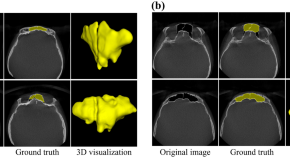
Automatic segmentation and classification of frontal sinuses for sex determination from CBCT scans using a two-stage anatomy-guided attention network
- Renan Lucio Berbel da Silva
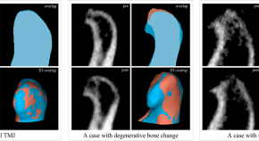
A study on volumetric change of mandibular condyles with osteoarthritis using cone-beam computed tomography
- Chang-Ki Min
- Kyoung-A Kim
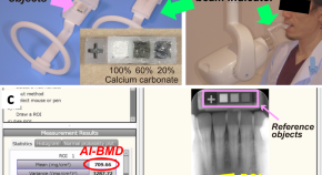
Sex differences and age-related changes in the mandibular alveolar bone mineral density using a computer-aided measurement system for intraoral radiography
- Ryutaro Ono
- Akitoshi Katsumata
- Narisato Kanamura
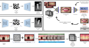
Generative artificial intelligence: synthetic datasets in dentistry
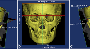

Facial asymmetry of the hard and soft tissues in skeletal Class I, II, and III patients
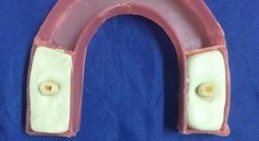
Introducing a new auto edge detection technique capable of revealing cervical root resorption in CBCT scans with pronounced metallic artifacts
- Negar Khosravifard
- Bardia Vadiati Saberi
- Mohammad Ebrahim Ghaffari
News and Comment
Artificial intelligence and dental panoramic radiographs: where are we now.
- Scott Webster
- Jacqueline Fraser
Does the use of antimicrobials in different periodontal treatment strategies result in better treatment outcomes? – A radiographic analysis
- Ryan McSorley
Artificial intelligence: is it more accurate than endodontists in root canal therapy?
- Mohammed Murad
- Faleh Tamimi
Apical periodontitis and autoimmune diseases—should we be screening patients prior to therapy?
The arthur prophet memorial lecture.
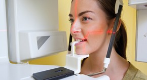
New regulations on X-ray use: likely implications for dental practices
Professor Keith Horner, University of Manchester, Co-editor of FGDP(UK)'s Selection criteria for dental radiography , has reviewed the draft Ionising Radiation Regulations 2017 and draft Ionising Radiation (Medical Exposure) Regulations 2018 and what they mean for dental practices.
- Keith Horner
Quick links
- Explore articles by subject
- Guide to authors
- Editorial policies

Theme : Emerging trends in Oral Medicine and Radiology

PAPER PRESENTATION
Click Here to Submit
POSTER PRESENTATION
Guidelines for abstract submission.
- Faculties, Post-graduate students and private practitioners belonging to the specialty of Oral Medicine and Radiology are eligible to participate in the event.
- Registered participant can present only one presentation, either paper or poster.
- The last date for abstract submission will be final and will not be modified.
- It is mandatory that each author can present only one paper. No co-authors allowed.
- Two authors (one author and one co-author) can present a poster.
- Word limit for abstract is 250 words.
- Structured abstract for original research and non-structured abstract for case report and review should be submitted.
- Abstracts for paper and poster should be submitted in their respective links only.
- The soft copy of the powerpoint presentation and the e-poster should be submitted beforehand along with the abstract via google forms provided in the website.
- Adherence to time limit and rules and regulations of scientific committee of the Conference is mandatory for all events.
- All presenters will be awarded the participation certificate for paper and poster presentation after their respective sessions.
- The results for the best paper/poster presentation will be announced during the valedictory function.
- The decision of the scientific committee will be final.
- TARA
- School of Dental Sciences
- Oral Medicine
Oral Medicine (Theses and Dissertations)
All of tara, this collection, date issued.
- Date of Publication
Search within this collection:
Recent Submissions
Oral leukoplakia - an investigation of its microbiome and of the association of clinical and microbial features with the degree of dysplasia , aspects of dose optimisation and diagnostic efficacy relating to dental cone beam computed tomography (cbct) , functional analysis of the tlo gene family in candida albicans and candida dubliniensis , molecular epidemiological typing of emerging methicillin-resistant staphylococcus aureus strains in the community, among livestock and in healthcare facilities in ireland, 2001-2015 , the role of hyphae and hypha-associated genes in the pathogenesis of candida dubliniensis .

Top 100 cited systematic reviews and meta-analyses in the major journals of oral and maxillofacial surgery : a bibliometric analysis
- Find this author on Google Scholar
- Find this author on PubMed
- Search for this author on this site
- ORCID record for Essam Ahmed Almoraissi
- For correspondence: [email protected]
- Info/History
- Preview PDF
The aim of this bibliometric research was to identify and analyze the top 100 cited systematic reviews in the field of oral and maxillofacial surgery. Using the Web of Science-database without restrictions on publication year or language, a bibliometric analysis was performed for the five major journals of oral and maxillofacial surgery: International Journal of Oral and Maxillofacial Surgery, Journal of Oral and Maxillofacial Surgery, Journal of Cranio-maxillofacial Surgery, British Journal of Oral & Maxillofacial Surgery and Oral Surgery Oral medicine Oral pathology Oral radiology. The most top-cited systematic review was published in 2015 with a total of 200 citations on survival and success rates of dental implants, consistent with the finding that “pre- and peri-implant surgery and dental implantology”, and “craniomaxillofacial deformities and cosmetic surgery” were the most frequently-cited topics (22% each). The International Journal of Oral and Maxillofacial Surgery and Journal of Oral and Maxillofacial Surgery displayed have got most citations in total and in average per publication. The outcome of this article can be used as a source of information not just for researchers but also for clinicians and students, and of which areas have a large impact on the field of oral and maxillofacial surgery but cannot reflect the quality of the included systematic reviews.
Competing Interest Statement
The authors have declared no competing interest.
Funding Statement
Author declarations.
I confirm all relevant ethical guidelines have been followed, and any necessary IRB and/or ethics committee approvals have been obtained.
The details of the IRB/oversight body that provided approval or exemption for the research described are given below:
not required
All necessary patient/participant consent has been obtained and the appropriate institutional forms have been archived.
I understand that all clinical trials and any other prospective interventional studies must be registered with an ICMJE-approved registry, such as ClinicalTrials.gov. I confirm that any such study reported in the manuscript has been registered and the trial registration ID is provided (note: if posting a prospective study registered retrospectively, please provide a statement in the trial ID field explaining why the study was not registered in advance).
I have followed all appropriate research reporting guidelines and uploaded the relevant EQUATOR Network research reporting checklist(s) and other pertinent material as supplementary files, if applicable.
Data Availability
View the discussion thread.
Thank you for your interest in spreading the word about medRxiv.
NOTE: Your email address is requested solely to identify you as the sender of this article.

Citation Manager Formats
- EndNote (tagged)
- EndNote 8 (xml)
- RefWorks Tagged
- Ref Manager
- Tweet Widget
- Facebook Like
- Google Plus One
Subject Area
- Dentistry and Oral Medicine
- Addiction Medicine (325)
- Allergy and Immunology (635)
- Anesthesia (168)
- Cardiovascular Medicine (2417)
- Dentistry and Oral Medicine (292)
- Dermatology (208)
- Emergency Medicine (382)
- Endocrinology (including Diabetes Mellitus and Metabolic Disease) (855)
- Epidemiology (11829)
- Forensic Medicine (10)
- Gastroenterology (705)
- Genetic and Genomic Medicine (3792)
- Geriatric Medicine (353)
- Health Economics (641)
- Health Informatics (2424)
- Health Policy (943)
- Health Systems and Quality Improvement (909)
- Hematology (344)
- HIV/AIDS (794)
- Infectious Diseases (except HIV/AIDS) (13370)
- Intensive Care and Critical Care Medicine (771)
- Medical Education (372)
- Medical Ethics (105)
- Nephrology (404)
- Neurology (3547)
- Nursing (200)
- Nutrition (531)
- Obstetrics and Gynecology (685)
- Occupational and Environmental Health (671)
- Oncology (1844)
- Ophthalmology (541)
- Orthopedics (223)
- Otolaryngology (287)
- Pain Medicine (234)
- Palliative Medicine (68)
- Pathology (450)
- Pediatrics (1044)
- Pharmacology and Therapeutics (429)
- Primary Care Research (425)
- Psychiatry and Clinical Psychology (3211)
- Public and Global Health (6212)
- Radiology and Imaging (1301)
- Rehabilitation Medicine and Physical Therapy (756)
- Respiratory Medicine (838)
- Rheumatology (381)
- Sexual and Reproductive Health (376)
- Sports Medicine (327)
- Surgery (409)
- Toxicology (51)
- Transplantation (174)
- Urology (148)
OPD Timing: 8.30am to 2.30pm (Sunday/National Holiday Off)
Admission BDS/MDS 2024-25 | Gallery | Contact Us
Paper and Poster Oral Medicine and Radiology
Best paper presentations.
- IAFO Conference, 2010- Chelioscopy.
- IAOMR conference, 2010- Facial palsy in poliomyelitis.
- OOO Symposium, 2011- Salivary mRNA in cancer Diagnostics.
- Rajasthan State IDA conference, 2012- Role of Dental Imaging on Forensic Dentistry.
Postgraduate student & interested under graduate students are encouraged to present atleast: one poster & one scientific paper presentation in various state well as in national conferences.
- Department Home
- Department Gallery
- Courses Offered
- Treatment Procedures
- Treatment Gallery
- Scientific and Other Academic Activities
- Awards and Achievements
Darshan Dental College and Hospital started in 1999 in the heart of Udaipur city of Rajasthan also famously known as the 'City of Lakes'. The college imparts education and training for 5 years leading to the degree of Bachelor of Dental Surgery (B.D.S) and Master in Dental Surgery (M.D.S) in all the specialties of dentistry.
Quick Links
- Departments
- Admission Inquiry
- News Updates
You've successfully sent the message!
Contact Info
- Ranakpur Road, Village-Loyara, Udaipur 313011 (Raj.)
- +91-8875677899, +91-9982682085
- [email protected]
- www.darshandentalcollege.org
- www.facebook.com/darshandental
- www.google.co.in/maps/darshandental
Copyright 2024 © All Rights Reserved
Designed by Aivon Solutions
- IAOMR - Indian Academy of Oral Medicine and Radiology was initiated in the year 1985.
- To endeavour to develop higher standard in teaching and practice of Oral Medicine, Oral Diagnosis, Maxillo-facial Radiology and Imaging Sciences.
- To promote Continuing Education, Research and Community Service to rural and urban population with grants, sponsorships and funds of the Academy.
- To revise the syllabus, curriculum, examination patterns for UG & PG courses as and when the need arises and recommend to the competent authorities to implement the same.
- To publish periodicals, journals, books etc., and to encourage the Academy members to pursue research and publish high quality scientific articles, and propagate knowledge of oral health care through different media.
IAOMR TIMES VOLUME 2 ISSUE 1
01/04/2024 ...
NATIONAL IAOMR PG CONVENTION 2024
24/08/2024 ...
IAOMR times - Fourth Issue(October-December 2023)
01/01/2024 ...
AERB Guidelines
...
Who is saying what
" I am thankful to the members for bestowing the honors and distinction by electing me the President of the Indian Academy of Oral Medicine and Radiology. It is your greatness and nobility that you have confidence in my capabilities. I take pride in conveying my thankfulness to the past Presidents and Office Bearers for their effort and endeavor in working hard to make immense progress and development in the specialty." Read more...
"It is an honour to serve as your Secretary at the Indian Academy of Oral Medicine and Radiology. I am committed to facilitating effective communication, promoting collaboration, and advancing our shared goals in oral medicine and radiology." Read more...
- Search Menu
- Sign in through your institution
- Volume 2024, Issue 6, June 2024 (In Progress)
- Volume 2024, Issue 5, May 2024
- Bariatric Surgery
- Breast Surgery
- Cardiothoracic Surgery
- Colorectal Surgery
- Colorectal Surgery, Upper GI Surgery
- Gynaecology
- Hepatobiliary Surgery
- Interventional Radiology
- Neurosurgery
- Ophthalmology
- Oral and Maxillofacial Surgery
- Otorhinolaryngology - Head & Neck Surgery
- Paediatric Surgery
- Plastic Surgery
- Transplant Surgery
- Trauma & Orthopaedic Surgery
- Upper GI Surgery
- Vascular Surgery
- Author Guidelines
- Submission Site
- Open Access
- Reasons to Submit
- About Journal of Surgical Case Reports
- Editorial Board
- Advertising and Corporate Services
- Journals Career Network
- Self-Archiving Policy
- Journals on Oxford Academic
- Books on Oxford Academic

Article Contents
- Introduction
- Case report
- Conflict of interest statement
- < Previous
Diagnosis of an unusual orbital abscess following sub-Tenon's steroid injection: a case report
- Article contents
- Figures & tables
- Supplementary Data
Isha Gupta, Elliott Moussa, Karun Motupally, Sharon Morris, Diagnosis of an unusual orbital abscess following sub-Tenon's steroid injection: a case report, Journal of Surgical Case Reports , Volume 2024, Issue 5, May 2024, rjae339, https://doi.org/10.1093/jscr/rjae339
- Permissions Icon Permissions
Orbital abscesses are caused by infection within or near the orbit and show obvious signs of pain, proptosis and raised inflammatory markers. Diagnosis is based on clinical features and radiological imaging, and requires early antibiotics and often surgical drainage to save vision. Sub-Tenon’s injections of triamcinolone acetonide (TA) have caused localized infections in previous reports, which have responded to therapeutic interventions. Here we report a case where a delayed presentation of an orbital abscess secondary to sub-Tenon’s TA for persistent post-operative cystoid macular oedema, without obvious signs of infection, rapidly progressed to cause orbital compartment syndrome. Despite treatment, the patient lost complete vision in the affected eye. This case discusses the rare and unusual cause of abscess formation and a diagnostic dilemma.
- triamcinolone acetonide
- diagnostic imaging
- orbital abscess
- orbital cyst
- episcleral block
- orbital compartment syndrome
Email alerts
Citing articles via, affiliations.
- Online ISSN 2042-8812
- Copyright © 2024 Oxford University Press and JSCR Publishing Ltd
- About Oxford Academic
- Publish journals with us
- University press partners
- What we publish
- New features
- Open access
- Institutional account management
- Rights and permissions
- Get help with access
- Accessibility
- Advertising
- Media enquiries
- Oxford University Press
- Oxford Languages
- University of Oxford
Oxford University Press is a department of the University of Oxford. It furthers the University's objective of excellence in research, scholarship, and education by publishing worldwide
- Copyright © 2024 Oxford University Press
- Cookie settings
- Cookie policy
- Privacy policy
- Legal notice
This Feature Is Available To Subscribers Only
Sign In or Create an Account
This PDF is available to Subscribers Only
For full access to this pdf, sign in to an existing account, or purchase an annual subscription.
Phone Numbers
Routine and emergency care.
Companion Animal Hospital in Ithaca, NY for cats, dogs, exotics, and wildlife
Equine and Nemo Farm Animal Hospitals in Ithaca, NY for horses and farm animals
Cornell Ruffian Equine Specialists, on Long Island for every horse
Ambulatory and Production Medicine for service on farms within 30 miles of Ithaca, NY
Animal Health Diagnostic Center New York State Veterinary Diagnostic Laboratory
General Information
Cornell University College of Veterinary Medicine Ithaca, New York 14853-6401

Anatomical discovery could help canine patients with liver function issues
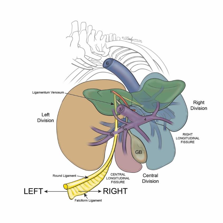
Portosystemic shunts are abnormal blood vessels that cause blood to bypass the liver, allowing toxins to build up. The shunts can develop in utero and lead to seizures, digestive problems, and stunted growth in puppies. Illustration: D. Sawchyn, MSMI, DVM, CMI of Sawchyn Medical Illustration, LLC (SMI)
Cornell veterinary radiologists have discovered anatomical details about a birth defect that commonly affects liver function in dogs – a finding that could aid in life-saving surgical repairs for many canine patients.
Portosystemic shunts are abnormal blood vessels that cause blood to bypass the liver, allowing toxins to build up. The shunts can develop in utero and lead to seizures, digestive problems, and stunted growth in puppies.
Radiology resident Nicholas Walsh, D.V.M.'19 worked with Peter V. Scrivani ’89, D.V.M. ’93, professor of radiology, to investigate the anatomical components of this defect. They published the results this month in the journal of Veterinary Radiology and Ultrasound.
“Through our study, we developed a concise and accurate way to describe these birth defects that clarifies our overall understanding of their anatomy,” Walsh explained. “Portosystemic shunts are a complex topic where anatomy plays a big role in our treatment planning. Our discoveries are exciting because they have practical clinical applications for patients.”
An astonishing recognition
The imaging team evaluated CT scans of this birth defect in 56 dogs from Cornell’s Companion Animal Hospital and the Schwarzman Animal Medical Center in New York City. The scans zero in specifically on intrahepatic portosystemic shunts – abnormal blood vessels often described as going through liver tissue, instead of around it. The study revealed that about half of these abnormal vessels are actually sandwiched between liver lobes, but on the outside of the liver.
“For a lot of us who have been performing CT for a long time, this was an astonishing recognition,” Scrivani said. “This new way of characterizing portosystemic shunts will help us provide really good diagnoses and communicate better with surgeons to make the most of novel surgical treatments for this defect.”
For his work on the paper, Walsh earned the Best Scientific Content award at the 18th Cornell University College of Veterinary Medicine Clinical Investigators’ Day, an opportunity for residents and interns to present their research to the veterinary community and practice their oral presentation skills.
Full circle
Cornell alumni and current faculty members collaborated on the study. Lauren D. Sawchyn, D.V.M. ’09, a board-certified veterinarian and medical illustrator, created the diagrams in the paper. Anthony Fischetti, D.V.M ‘01, head of radiology at the Schwarzman Animal Medical Center, contributed to the paper. And veterinarians from Cornell’s Department of Clinical Sciences and the Department of Biomedical Sciences helped the team to understand the anatomy of this defect.
“It was such a collaborative effort,” Walsh said. “This is why I love radiology. We are here to work with and support so many of the various disciplines in veterinary medicine.”
For Scrivani, who recently was promoted to full professor Cornell, this paper brings his career full circle. In 1993, he was a senior veterinary student at Cornell when he first learned about portosystemic shunt imaging.
“I did my senior seminar paper on the imaging of portosystemic shunts,” he said. “Back then, we were limited in the imaging technology available. Today, we have advanced CT angiograms that give us a very detailed pictures of the liver and allow us to see the blood vessels in excellent detail.”
Scrivani hopes the discoveries in this paper will improve the care for canine patients across the globe. “We’ve already gotten emails from people around the world who are excited to use our new classification system,” he said. “We’re hopeful it will make a difference for patients.”
Written by Sheri Hall
Recent News
Cornell University College of Veterinary Medicine recognizes newest graduates

CVM faculty, staff, awarded SUNY Chancellor’s Awards for Excellence

Wing skeleton evolution may be less restricted in small birds
Cornell immunology symposium -- june 3-5, 2024, educating generalists for adaptive expertise, summer lecture series: "how global fisheries connect us all—environmental change for health and well-being".

IMAGES
VIDEO
COMMENTS
Introduction. A thesis or dissertation, as some people would like to call it, is an integral part of the Radiology curriculum, be it MD, DNB, or DMRD. We have tried to aggregate radiology thesis topics from various sources for reference. Not everyone is interested in research, and writing a Radiology thesis can be daunting.
Oral Medicine & Radiology Oral medicine is the discipline in the dentistry which is used for diagnosis and non-surgical treatment of oral cavity and oral manifestations of systemic disease. Oral radiology is the branch of dentistry which dealing with use of x-rays, radioactive substances, and other forms of radiant energy in diagnosis and ...
Explore the latest full-text research PDFs, articles, conference papers, preprints and more on ORAL RADIOLOGY. Find methods information, sources, references or conduct a literature review on ORAL ...
Advancements in Telemedicine and Teleradiology: Pioneering Progress in Oral Medicine and Radiology. Journal of Indian Academy of Oral Medicine & Radiology. 35 (3):295-296, Jul-Sep 2023.
Virtual Reality and Augmented Reality in Oral Medicine. Conventionally, oral cavity examinations and clinical diagnostic investigations of oral lesions were either explained orally or with visual presentations. The oral medicine residents are expected to take a proper clinical history of the patient with a thorough oral cavity examination.
Feature papers represent the most advanced research with significant potential for high impact in the field. ... This Special Issue aims to provide an update on the latest research and advances in oral medicine that can help improve clinical decision-making and impact the quality of life of our patients. Topics of interest include: Oral cancer ...
Letter to the editor: Magnetic resonance imaging-based radiomics and deep learning models for predicting lymph node metastasis of squamous cell carcinoma of the tongue. Zhe Hu, Zhikang Tian, Xi Wei, Yueqin Chen. In Press, Journal Pre-proof, Available online 3 May 2024. View PDF.
Overview. Oral Radiology is a peer-reviewed English-language journal that publishes cutting-edge research papers and reviews in the fields of diagnostic imaging of the head and neck and all related fields. The official journal of the Japanese Society for Oral and Maxillofacial Radiology and the Asian Academy of Oral and Maxillofacial Radiology.
A selection of abstracts of clinically relevant papers from other journals. The abstracts on this page have been chosen and edited by John R. Radford. Research Highlights 07 Apr 2017 British ...
Therapeutic Evaluation of 5% Topical Amlexanox Paste and 2% Curcumin Oral Gel in the Management of Recurrent Aphthous Stomatitis- A Randomized Clinical Trial. Gauthaman, Jeevitha; Ganesan, Anuradha. Journal of Indian Academy of Oral Medicine and Radiology. 34 (1):17-21, Jan-Mar 2022. Abstract.
Advances in diagnostic oral medicine are aimed at reducing. the morbidity and mortality associated with oral diseases. For e xample, despite numerous advances in treatment, the 5-year survival ...
Professor Keith Horner, University of Manchester, Co-editor of FGDP(UK)'s Selection criteria for dental radiography, has reviewed the draft Ionising Radiation Regulations 2017 and draft Ionising ...
Correlation of the Mandibular Inferior Cortical Index and Panoramic Mandibular Index on Digital Panoramic Radiographs with Total Serum Calcium and Maximum Hand-Grip Strength in the Elderly - A Prospective Study. Journal of Indian Academy of Oral Medicine and Radiology. 34 (4):452-455, Oct-Dec 2022.
Introduction: The Oral Surgery Specialized Graduate Degree (Diplôme d'Études Spécialisées en Chirurgie Orale, DESCO) was created in France in 2011. The purpose of this study was to assess ...
Faculties, Post-graduate students and private practitioners belonging to the specialty of Oral Medicine and Radiology are eligible to participate in the event. Registered participant can present only one presentation, either paper or poster. The last date for abstract submission will be final and will not be modified.
ORAL MEDICINE & RADIOLOGY SYLLABUS 1. Methods of clinical diagnosis of oral and systemic diseases as applicable to oral tissue ... Paper presentation -2, poster presentation 2, article publication 2 19. Library dissertation to be submitted before end of 3rd term, final thesis as per Instructions of student section . I. ORAL MEDICINE: 1. A) Case ...
Oral leukoplakia - an investigation of its microbiome and of the association of clinical and microbial features with the degree of dysplasia . Galvin, Sheila. Background: With growing evidence of a shift in the oral microbiome associated with oral squamous cell carcinoma, it was hypothesised that the microbiome of oral leukoplakia (OLK), the ...
The aim of this bibliometric research was to identify and analyze the top 100 cited systematic reviews in the field of oral and maxillofacial surgery. Using the Web of Science-database without restrictions on publication year or language, a bibliometric analysis was performed for the five major journals of oral and maxillofacial surgery: International Journal of Oral and Maxillofacial Surgery ...
Best paper presentations. IAFO Conference, 2010- Chelioscopy. IAOMR conference, 2010- Facial palsy in poliomyelitis. OOO Symposium, 2011- Salivary mRNA in cancer Diagnostics. Rajasthan State IDA conference, 2012- Role of Dental Imaging on Forensic Dentistry. Postgraduate student & interested under graduate students are encouraged to present ...
IAOMR. IAOMR - Indian Academy of Oral Medicine and Radiology was initiated in the year 1985. To endeavour to develop higher standard in teaching and practice of Oral Medicine, Oral Diagnosis, Maxillo-facial Radiology and Imaging Sciences. To promote Continuing Education, Research and Community Service to rural and urban population with grants ...
An Oral Medicine specialist is a "Physician of the mouth". The speciality manages oral mucosal diseases, salivary gland diseases. Oral cancer screening, tobacco cessation initiatives and management of disease using intralesional injections are some aspects of the professional training programme. Oral Radiology is an integral part of Oral ...
Dissertation topics and names of Guides and students S.N o NAME OF P.G. NAME OF GUIDE YEAR THESIS TOPIC 1. Dr. Upendra Malik Dr. M Srinivasa Raju 2006 Management Of Oral Lichen Planus With Topical Tacrolimus 2 Dr. Nitin Nigam Dr. M Srinivasa Raju 2006 Prevalence Of Oral Submucous Fibrosis Among Gutkha And Areca Nut Chewers In Moradabad City
The Department has adequate space for oral diagnosis and radiology and functions in two sections:- In the ORAL MEDICINE clinic, all the patients who come to the dental college and hospital are examined. Diagnosis of diseases with scientific methods and laboratory investigations is done. All cases with oromucosal lesions are treated with various ...
Prostate cancer lung metastasis represents a clinical conundrum due to its implications for advanced disease progression and the complexities it introduces in treatment planning. As the disease progresses to distant sites such as the lung, the clinical management becomes increasingly intricate, requiring tailored therapeutic strategies to address the unique characteristics of metastatic ...
Here we report a case where a delayed presentation of an orbital abscess secondary to sub-Tenon's TA for persistent post-operative cystoid macular oedema, without obvious signs of infection, rapidly progressed to cause orbital compartment syndrome. Despite treatment, the patient lost complete vision in the affected eye.
For his work on the paper, Walsh earned the Best Scientific Content award at the 18th Cornell University College of Veterinary Medicine Clinical Investigators' Day, an opportunity for residents and interns to present their research to the veterinary community and practice their oral presentation skills. Full circle