- TPC and eLearning
- What's NEW at TPC?
- Read Watch Interact
- Practice Review Test
- Teacher-Tools
- Request a Demo
- Get A Quote
- Subscription Selection
- Seat Calculator
- Ad Free Account
- Edit Profile Settings
- Metric Conversions Questions
- Metric System Questions
- Metric Estimation Questions
- Significant Digits Questions
- Proportional Reasoning
- Acceleration
- Distance-Displacement
- Dots and Graphs
- Graph That Motion
- Match That Graph
- Name That Motion
- Motion Diagrams
- Pos'n Time Graphs Numerical
- Pos'n Time Graphs Conceptual
- Up And Down - Questions
- Balanced vs. Unbalanced Forces
- Change of State
- Force and Motion
- Mass and Weight
- Match That Free-Body Diagram
- Net Force (and Acceleration) Ranking Tasks
- Newton's Second Law
- Normal Force Card Sort
- Recognizing Forces
- Air Resistance and Skydiving
- Solve It! with Newton's Second Law
- Which One Doesn't Belong?
- Component Addition Questions
- Head-to-Tail Vector Addition
- Projectile Mathematics
- Trajectory - Angle Launched Projectiles
- Trajectory - Horizontally Launched Projectiles
- Vector Addition
- Vector Direction
- Which One Doesn't Belong? Projectile Motion
- Forces in 2-Dimensions
- Being Impulsive About Momentum
- Explosions - Law Breakers
- Hit and Stick Collisions - Law Breakers
- Case Studies: Impulse and Force
- Impulse-Momentum Change Table
- Keeping Track of Momentum - Hit and Stick
- Keeping Track of Momentum - Hit and Bounce
- What's Up (and Down) with KE and PE?
- Energy Conservation Questions
- Energy Dissipation Questions
- Energy Ranking Tasks
- LOL Charts (a.k.a., Energy Bar Charts)
- Match That Bar Chart
- Words and Charts Questions
- Name That Energy
- Stepping Up with PE and KE Questions
- Case Studies - Circular Motion
- Circular Logic
- Forces and Free-Body Diagrams in Circular Motion
- Gravitational Field Strength
- Universal Gravitation
- Angular Position and Displacement
- Linear and Angular Velocity
- Angular Acceleration
- Rotational Inertia
- Balanced vs. Unbalanced Torques
- Getting a Handle on Torque
- Torque-ing About Rotation
- Properties of Matter
- Fluid Pressure
- Buoyant Force
- Sinking, Floating, and Hanging
- Pascal's Principle
- Flow Velocity
- Bernoulli's Principle
- Balloon Interactions
- Charge and Charging
- Charge Interactions
- Charging by Induction
- Conductors and Insulators
- Coulombs Law
- Electric Field
- Electric Field Intensity
- Polarization
- Case Studies: Electric Power
- Know Your Potential
- Light Bulb Anatomy
- I = ∆V/R Equations as a Guide to Thinking
- Parallel Circuits - ∆V = I•R Calculations
- Resistance Ranking Tasks
- Series Circuits - ∆V = I•R Calculations
- Series vs. Parallel Circuits
- Equivalent Resistance
- Period and Frequency of a Pendulum
- Pendulum Motion: Velocity and Force
- Energy of a Pendulum
- Period and Frequency of a Mass on a Spring
- Horizontal Springs: Velocity and Force
- Vertical Springs: Velocity and Force
- Energy of a Mass on a Spring
- Decibel Scale
- Frequency and Period
- Closed-End Air Columns
- Name That Harmonic: Strings
- Rocking the Boat
- Wave Basics
- Matching Pairs: Wave Characteristics
- Wave Interference
- Waves - Case Studies
- Color Addition and Subtraction
- Color Filters
- If This, Then That: Color Subtraction
- Light Intensity
- Color Pigments
- Converging Lenses
- Curved Mirror Images
- Law of Reflection
- Refraction and Lenses
- Total Internal Reflection
- Who Can See Who?
- Lab Equipment
- Lab Procedures
- Formulas and Atom Counting
- Atomic Models
- Bond Polarity
- Entropy Questions
- Cell Voltage Questions
- Heat of Formation Questions
- Reduction Potential Questions
- Oxidation States Questions
- Measuring the Quantity of Heat
- Hess's Law
- Oxidation-Reduction Questions
- Galvanic Cells Questions
- Thermal Stoichiometry
- Molecular Polarity
- Quantum Mechanics
- Balancing Chemical Equations
- Bronsted-Lowry Model of Acids and Bases
- Classification of Matter
- Collision Model of Reaction Rates
- Density Ranking Tasks
- Dissociation Reactions
- Complete Electron Configurations
- Elemental Measures
- Enthalpy Change Questions
- Equilibrium Concept
- Equilibrium Constant Expression
- Equilibrium Calculations - Questions
- Equilibrium ICE Table
- Intermolecular Forces Questions
- Ionic Bonding
- Lewis Electron Dot Structures
- Limiting Reactants
- Line Spectra Questions
- Mass Stoichiometry
- Measurement and Numbers
- Metals, Nonmetals, and Metalloids
- Metric Estimations
- Metric System
- Molarity Ranking Tasks
- Mole Conversions
- Name That Element
- Names to Formulas
- Names to Formulas 2
- Nuclear Decay
- Particles, Words, and Formulas
- Periodic Trends
- Precipitation Reactions and Net Ionic Equations
- Pressure Concepts
- Pressure-Temperature Gas Law
- Pressure-Volume Gas Law
- Chemical Reaction Types
- Significant Digits and Measurement
- States Of Matter Exercise
- Stoichiometry Law Breakers
- Stoichiometry - Math Relationships
- Subatomic Particles
- Spontaneity and Driving Forces
- Gibbs Free Energy
- Volume-Temperature Gas Law
- Acid-Base Properties
- Energy and Chemical Reactions
- Chemical and Physical Properties
- Valence Shell Electron Pair Repulsion Theory
- Writing Balanced Chemical Equations
- Mission CG1
- Mission CG10
- Mission CG2
- Mission CG3
- Mission CG4
- Mission CG5
- Mission CG6
- Mission CG7
- Mission CG8
- Mission CG9
- Mission EC1
- Mission EC10
- Mission EC11
- Mission EC12
- Mission EC2
- Mission EC3
- Mission EC4
- Mission EC5
- Mission EC6
- Mission EC7
- Mission EC8
- Mission EC9
- Mission RL1
- Mission RL2
- Mission RL3
- Mission RL4
- Mission RL5
- Mission RL6
- Mission KG7
- Mission RL8
- Mission KG9
- Mission RL10
- Mission RL11
- Mission RM1
- Mission RM2
- Mission RM3
- Mission RM4
- Mission RM5
- Mission RM6
- Mission RM8
- Mission RM10
- Mission LC1
- Mission RM11
- Mission LC2
- Mission LC3
- Mission LC4
- Mission LC5
- Mission LC6
- Mission LC8
- Mission SM1
- Mission SM2
- Mission SM3
- Mission SM4
- Mission SM5
- Mission SM6
- Mission SM8
- Mission SM10
- Mission KG10
- Mission SM11
- Mission KG2
- Mission KG3
- Mission KG4
- Mission KG5
- Mission KG6
- Mission KG8
- Mission KG11
- Mission F2D1
- Mission F2D2
- Mission F2D3
- Mission F2D4
- Mission F2D5
- Mission F2D6
- Mission KC1
- Mission KC2
- Mission KC3
- Mission KC4
- Mission KC5
- Mission KC6
- Mission KC7
- Mission KC8
- Mission AAA
- Mission SM9
- Mission LC7
- Mission LC9
- Mission NL1
- Mission NL2
- Mission NL3
- Mission NL4
- Mission NL5
- Mission NL6
- Mission NL7
- Mission NL8
- Mission NL9
- Mission NL10
- Mission NL11
- Mission NL12
- Mission MC1
- Mission MC10
- Mission MC2
- Mission MC3
- Mission MC4
- Mission MC5
- Mission MC6
- Mission MC7
- Mission MC8
- Mission MC9
- Mission RM7
- Mission RM9
- Mission RL7
- Mission RL9
- Mission SM7
- Mission SE1
- Mission SE10
- Mission SE11
- Mission SE12
- Mission SE2
- Mission SE3
- Mission SE4
- Mission SE5
- Mission SE6
- Mission SE7
- Mission SE8
- Mission SE9
- Mission VP1
- Mission VP10
- Mission VP2
- Mission VP3
- Mission VP4
- Mission VP5
- Mission VP6
- Mission VP7
- Mission VP8
- Mission VP9
- Mission WM1
- Mission WM2
- Mission WM3
- Mission WM4
- Mission WM5
- Mission WM6
- Mission WM7
- Mission WM8
- Mission WE1
- Mission WE10
- Mission WE2
- Mission WE3
- Mission WE4
- Mission WE5
- Mission WE6
- Mission WE7
- Mission WE8
- Mission WE9
- Vector Walk Interactive
- Name That Motion Interactive
- Kinematic Graphing 1 Concept Checker
- Kinematic Graphing 2 Concept Checker
- Graph That Motion Interactive
- Two Stage Rocket Interactive
- Rocket Sled Concept Checker
- Force Concept Checker
- Free-Body Diagrams Concept Checker
- Free-Body Diagrams The Sequel Concept Checker
- Skydiving Concept Checker
- Elevator Ride Concept Checker
- Vector Addition Concept Checker
- Vector Walk in Two Dimensions Interactive
- Name That Vector Interactive
- River Boat Simulator Concept Checker
- Projectile Simulator 2 Concept Checker
- Projectile Simulator 3 Concept Checker
- Hit the Target Interactive
- Turd the Target 1 Interactive
- Turd the Target 2 Interactive
- Balance It Interactive
- Go For The Gold Interactive
- Egg Drop Concept Checker
- Fish Catch Concept Checker
- Exploding Carts Concept Checker
- Collision Carts - Inelastic Collisions Concept Checker
- Its All Uphill Concept Checker
- Stopping Distance Concept Checker
- Chart That Motion Interactive
- Roller Coaster Model Concept Checker
- Uniform Circular Motion Concept Checker
- Horizontal Circle Simulation Concept Checker
- Vertical Circle Simulation Concept Checker
- Race Track Concept Checker
- Gravitational Fields Concept Checker
- Orbital Motion Concept Checker
- Angular Acceleration Concept Checker
- Balance Beam Concept Checker
- Torque Balancer Concept Checker
- Aluminum Can Polarization Concept Checker
- Charging Concept Checker
- Name That Charge Simulation
- Coulomb's Law Concept Checker
- Electric Field Lines Concept Checker
- Put the Charge in the Goal Concept Checker
- Circuit Builder Concept Checker (Series Circuits)
- Circuit Builder Concept Checker (Parallel Circuits)
- Circuit Builder Concept Checker (∆V-I-R)
- Circuit Builder Concept Checker (Voltage Drop)
- Equivalent Resistance Interactive
- Pendulum Motion Simulation Concept Checker
- Mass on a Spring Simulation Concept Checker
- Particle Wave Simulation Concept Checker
- Boundary Behavior Simulation Concept Checker
- Slinky Wave Simulator Concept Checker
- Simple Wave Simulator Concept Checker
- Wave Addition Simulation Concept Checker
- Standing Wave Maker Simulation Concept Checker
- Color Addition Concept Checker
- Painting With CMY Concept Checker
- Stage Lighting Concept Checker
- Filtering Away Concept Checker
- InterferencePatterns Concept Checker
- Young's Experiment Interactive
- Plane Mirror Images Interactive
- Who Can See Who Concept Checker
- Optics Bench (Mirrors) Concept Checker
- Name That Image (Mirrors) Interactive
- Refraction Concept Checker
- Total Internal Reflection Concept Checker
- Optics Bench (Lenses) Concept Checker
- Kinematics Preview
- Velocity Time Graphs Preview
- Moving Cart on an Inclined Plane Preview
- Stopping Distance Preview
- Cart, Bricks, and Bands Preview
- Fan Cart Study Preview
- Friction Preview
- Coffee Filter Lab Preview
- Friction, Speed, and Stopping Distance Preview
- Up and Down Preview
- Projectile Range Preview
- Ballistics Preview
- Juggling Preview
- Marshmallow Launcher Preview
- Air Bag Safety Preview
- Colliding Carts Preview
- Collisions Preview
- Engineering Safer Helmets Preview
- Push the Plow Preview
- Its All Uphill Preview
- Energy on an Incline Preview
- Modeling Roller Coasters Preview
- Hot Wheels Stopping Distance Preview
- Ball Bat Collision Preview
- Energy in Fields Preview
- Weightlessness Training Preview
- Roller Coaster Loops Preview
- Universal Gravitation Preview
- Keplers Laws Preview
- Kepler's Third Law Preview
- Charge Interactions Preview
- Sticky Tape Experiments Preview
- Wire Gauge Preview
- Voltage, Current, and Resistance Preview
- Light Bulb Resistance Preview
- Series and Parallel Circuits Preview
- Thermal Equilibrium Preview
- Linear Expansion Preview
- Heating Curves Preview
- Electricity and Magnetism - Part 1 Preview
- Electricity and Magnetism - Part 2 Preview
- Vibrating Mass on a Spring Preview
- Period of a Pendulum Preview
- Wave Speed Preview
- Slinky-Experiments Preview
- Standing Waves in a Rope Preview
- Sound as a Pressure Wave Preview
- DeciBel Scale Preview
- DeciBels, Phons, and Sones Preview
- Sound of Music Preview
- Shedding Light on Light Bulbs Preview
- Models of Light Preview
- Electromagnetic Radiation Preview
- Electromagnetic Spectrum Preview
- EM Wave Communication Preview
- Digitized Data Preview
- Light Intensity Preview
- Concave Mirrors Preview
- Object Image Relations Preview
- Snells Law Preview
- Reflection vs. Transmission Preview
- Magnification Lab Preview
- Reactivity Preview
- Ions and the Periodic Table Preview
- Periodic Trends Preview
- Chemical Reactions Preview
- Intermolecular Forces Preview
- Melting Points and Boiling Points Preview
- Bond Energy and Reactions Preview
- Reaction Rates Preview
- Ammonia Factory Preview
- Stoichiometry Preview
- Nuclear Chemistry Preview
- Gaining Teacher Access
- Task Tracker Directions
- Conceptual Physics Course
- On-Level Physics Course
- Honors Physics Course
- Chemistry Concept Builders
- All Chemistry Resources
- Users Voice
- Tasks and Classes
- Webinars and Trainings
- Subscription
- Subscription Locator
- 1-D Kinematics
- Newton's Laws
- Vectors - Motion and Forces in Two Dimensions
- Momentum and Its Conservation
- Work and Energy
- Circular Motion and Satellite Motion
- Thermal Physics
- Static Electricity
- Electric Circuits
- Vibrations and Waves
- Sound Waves and Music
- Light and Color
- Reflection and Mirrors
- Measurement and Calculations
- Elements, Atoms, and Ions
- About the Physics Interactives
- Task Tracker
- Usage Policy
- Newtons Laws
- Vectors and Projectiles
- Forces in 2D
- Momentum and Collisions
- Circular and Satellite Motion
- Balance and Rotation
- Electromagnetism
- Waves and Sound
- Atomic Physics
- Forces in Two Dimensions
- Work, Energy, and Power
- Circular Motion and Gravitation
- Sound Waves
- 1-Dimensional Kinematics
- Circular, Satellite, and Rotational Motion
- Einstein's Theory of Special Relativity
- Waves, Sound and Light
- QuickTime Movies
- About the Concept Builders
- Pricing For Schools
- Directions for Version 2
- Measurement and Units
- Relationships and Graphs
- Rotation and Balance
- Vibrational Motion
- Reflection and Refraction
- Teacher Accounts
- Kinematic Concepts
- Kinematic Graphing
- Wave Motion
- Sound and Music
- About CalcPad
- 1D Kinematics
- Vectors and Forces in 2D
- Simple Harmonic Motion
- Rotational Kinematics
- Rotation and Torque
- Rotational Dynamics
- Electric Fields, Potential, and Capacitance
- Transient RC Circuits
- Light Waves
- Units and Measurement
- Stoichiometry
- Molarity and Solutions
- Thermal Chemistry
- Acids and Bases
- Kinetics and Equilibrium
- Solution Equilibria
- Oxidation-Reduction
- Nuclear Chemistry
- Newton's Laws of Motion
- Work and Energy Packet
- Static Electricity Review
- NGSS Alignments
- 1D-Kinematics
- Projectiles
- Circular Motion
- Magnetism and Electromagnetism
- Graphing Practice
- About the ACT
- ACT Preparation
- For Teachers
- Other Resources
- Solutions Guide
- Solutions Guide Digital Download
- Motion in One Dimension
- Work, Energy and Power
- Chemistry of Matter
- Measurement and the Metric System
- Names and Formulas
- Algebra Based On-Level Physics
- Honors Physics
- Conceptual Physics
- Other Tools
- Frequently Asked Questions
- Purchasing the Download
- Purchasing the Digital Download
- About the NGSS Corner
- NGSS Search
- Force and Motion DCIs - High School
- Energy DCIs - High School
- Wave Applications DCIs - High School
- Force and Motion PEs - High School
- Energy PEs - High School
- Wave Applications PEs - High School
- Crosscutting Concepts
- The Practices
- Physics Topics
- NGSS Corner: Activity List
- NGSS Corner: Infographics
- About the Toolkits
- Position-Velocity-Acceleration
- Position-Time Graphs
- Velocity-Time Graphs
- Newton's First Law
- Newton's Second Law
- Newton's Third Law
- Terminal Velocity
- Projectile Motion
- Forces in 2 Dimensions
- Impulse and Momentum Change
- Momentum Conservation
- Work-Energy Fundamentals
- Work-Energy Relationship
- Roller Coaster Physics
- Satellite Motion
- Electric Fields
- Circuit Concepts
- Series Circuits
- Parallel Circuits
- Describing-Waves
- Wave Behavior Toolkit
- Standing Wave Patterns
- Resonating Air Columns
- Wave Model of Light
- Plane Mirrors
- Curved Mirrors
- Teacher Guide
- Using Lab Notebooks
- Current Electricity
- Light Waves and Color
- Reflection and Ray Model of Light
- Refraction and Ray Model of Light
- Teacher Resources
- Subscriptions

- Newton's Laws
- Einstein's Theory of Special Relativity
- About Concept Checkers
- School Pricing
- Newton's Laws of Motion
- Newton's First Law
- Newton's Third Law

Resonance and Air Columns - Complete Toolkit
- To describe how one object that vibrates at the same natural frequency of another object can force that second object into resonant vibrations and to identify and discuss several examples of such resonance phenomenon.
- To associate a resonating object with a standing wave pattern and to examine such patterns and identify their nodes and antinodes.
- To define fundamental frequency and to mathematically relate the fundamental frequency to the frequency of the various harmonics of a vibrating object.
- To draw the standing wave patterns for the various harmonics of an open-end and a closed-end air column and to relate the length of the column to the wavelength of the standing wave patterns.
- To use the speed-wavelength-frequency relationship to mathematically analyze a standing wave situation and relate the frequency of the harmonic to the length of the air column and the speed of sound waves within the air column.
Readings from The Physics Classroom Tutorial
- The Physics Classroom Tutorial, Sound Waves and Music Chapter, Lesson 4
- The Physics Classroom Tutorial, Sound Waves and Music Chapter, Lesson 5
Interactive Simulations
Video and Animations
Labs and Investigations
- The Physics Classroom, The Laboratory, Vibrating Spring Lab Students use a spring, a mechanical oscillator, and a frequency generator to create longitudinal standing waves in a spring and explore the relationship between the frequency and the spacing between adjacent nodes.
- The Physics Classroom, The Laboratory, Nodes and Antinodes Lab Students use a spring, a mechanical oscillator, and a frequency generator to create longitudinal standing waves in a spring and explore the relationship between the frequency and the number of nodes and antinodes present along the length of the spring. Link for Lab #1 and #2: http://www.physicsclassroom.com/lab/waves/Wlabs.cfm
- The Physics Classroom, The Laboratory, Closed End Air Column Lab Students use a tuning fork and a closed end air column to determine the speed of sound in air.
- The Physics Classroom, The Laboratory, Open-End Air Column Lab Students use an audio speaker driven by a frequency generator and an open end air column to determine the speed of sound in air. Link for Labs #3 and #4: http://www.physicsclassroom.com/lab/sound/Slabs.cfm
Demonstration Ideas
Minds On Physics Internet Modules:
- Sound and Music, Ass’t SM5 - Resonance and Standing Wave Patterns
- Sound and Music, Ass’t SM8 - Harmonics for Open-End Air Columns
- Sound and Music, Ass’t SM9 - Mathematics for Open-End Air Columns
- Sound and Music, Ass’t SM10 - Harmonics for Closed-End Air Columns
- Sound and Music, Ass’t SM11 - Mathematics for Closed -End Air Columns
Concept Building Exercises:
- The Curriculum Corner, Sound and Music, Resonance
- The Curriculum Corner, Sound and Music, Resonance and Open-End Air Columns
- The Curriculum Corner, Sound and Music, Resonance and Closed-End Air Columns
Problem-Solving Exercises:
- The Calculator Pad, Sound and Music, Problems #21 - #31 Link: http://www.physicsclassroom.com/calcpad/sound
Science Reasoning Activities:
- The Sound of Music Link: http://www.physicsclassroom.com/reasoning/sound
Cool Stuff Worth Showing:
Real Life Connections:
- HS-PS4-1 : The wavelength and frequency of a wave are related to one another by the speed of travel of the wave, which depends on the type of wave and the medium through which it is passing.
- Mathematical representations are needed to identify some patterns.
- Cause and effect relationships can be suggested and predicted for complex natural and human designed systems by examining what is known about smaller scale mechanisms within the system.
- Algebraic thinking is used to examine scientific data and predict the effect of a change in one variable on another (e.g., linear growth vs. exponential growth).
- Models (e.g., physical, mathematical, computer models) can be used to simulate systems and interactions—including energy, matter, and information flows—within and between systems at different scales.
- When investigating or describing a system, the boundaries and initial conditions of the system need to be defined and their inputs and outputs analyzed and described using models.
- Develop and/or use multiple types of models to provide mechanistic accounts of phenomena
- Develop and/or use a computational model to generate data to support explanations, predict phenomena, and analyze systems.
- Select appropriate tools to collect, record, analyze, and evaluate data.
- Make directional hypotheses that specify what happens to a dependent variable when an independent variable is manipulated.
- Apply concepts of statistics and probability (including determining function fits to data, slope, intercept, and correlation coefficient for linear fits) to scientific and engineering questions and problems, using digital tools when feasible.
- Use mathematical, computational, and/or algorithmic representations of phenomena or design solutions to describe and/or support claims and/or explanations.
- Apply techniques of algebra and functions to represent and solve scientific and engineering problems.
- Make and defend a claim based on evidence about the natural world or the effectiveness of a design solution that reflects scientific knowledge and student-generated evidence.
Demos: 4B-11 Resonance in Boxes with Tuning Forks

Add to Cart | View Cart
- Academic Programs
- Undergraduate
- Future Students
Department of Physics and Astronomy, 525 Northwestern Avenue, West Lafayette, IN 47907-2036 • Phone: (765) 494-3000 • Fax: (765) 494-0706
Copyright © 2024 Purdue University | An equal access/equal opportunity university | Copyright Complaints | DOE Degree Scorecards
Trouble with this page? Accessibility issues ? Please contact the College of Science .
14.4 Sound Interference and Resonance
Section learning objectives.
By the end of this section, you will be able to do the following:
- Describe resonance and beats
- Define fundamental frequency and harmonic series
- Contrast an open-pipe and closed-pipe resonator
- Solve problems involving harmonic series and beat frequency
Teacher Support
The learning objectives in this section will help your students master the following standards:
- (D) investigate behaviors of waves, including reflection, refraction, diffraction, interference, resonance, and the Doppler effect.
In addition, the High School Physics Laboratory Manual addresses content in this section in the lab titled: Sound Waves, as well as the following standards:
Section Key Terms
| beat | beat frequency | damping | fundamental | harmonics |
| natural frequency | overtones | resonance | resonate |
[BL] Before the start of this section, it would be useful to review the properties of sound waves and how they are related to each other, standing waves, superposition and interference of waves.
Resonance and Beats
Sit in front of a piano sometime and sing a loud brief note at it while pushing down on the sustain pedal. It will sing the same note back at you—the strings that have the same frequencies as your voice, are resonating in response to the forces from the sound waves that you sent to them. This is a good example of the fact that objects—in this case, piano strings—can be forced to oscillate but oscillate best at their natural frequency.
A driving force (such as your voice in the example) puts energy into a system at a certain frequency, which is not necessarily the same as the natural frequency of the system. Over time the energy dissipates, and the amplitude gradually reduces to zero- this is called damping . The natural frequency is the frequency at which a system would oscillate if there were no driving and no damping force. The phenomenon of driving a system with a frequency equal to its natural frequency is called resonance , and a system being driven at its natural frequency is said to resonate .
Most of us have played with toys where an object bobs up and down on an elastic band, something like the paddle ball suspended from a finger in Figure 14.18 . At first you hold your finger steady, and the ball bounces up and down with a small amount of damping. If you move your finger up and down slowly, the ball will follow along without bouncing much on its own. As you increase the frequency at which you move your finger up and down, the ball will respond by oscillating with increasing amplitude. When you drive the ball at its natural frequency, the ball’s oscillations increase in amplitude with each oscillation for as long as you drive it. As the driving frequency gets progressively higher than the resonant or natural frequency, the amplitude of the oscillations becomes smaller, until the oscillations nearly disappear and your finger simply moves up and down with little effect on the ball.
Another example is that when you tune a radio, you adjust its resonant frequency so that it oscillates only at the desired station’s broadcast (driving) frequency. Also, a child on a swing is driven (pushed) by a parent at the swing’s natural frequency to reach the maximum amplitude (height). In all of these cases, the efficiency of energy transfer from the driving force into the oscillator is best at resonance.
[BL] [OL] [AL] Tuning forks and pipes may be used to demonstrate the concept of resonance. Use any pipe or tube closed at one end. Fix it so that it stands upright with the open end on top. Choose a tuning fork and strike it to make it vibrate. Place it near the mouth of the pipe and hear the sound. Now fill the pipe with some water and repeat. The changing water level changes the length of the resonating air column. Continue doing this. When a certain length is obtained, the sound of the tuning fork will resonate through the column.
All sound resonances are due to constructive and destructive interference. Only the resonant frequencies interfere constructively to form standing waves, while others interfere destructively and are absent. From the toot made by blowing over a bottle to the recognizability of a great singer’s voice, resonance and standing waves play a vital role in sound.
Interference happens to all types of waves, including sound waves. In fact, one way to support that something is a wave is to observe interference effects. Figure 14.19 shows a set of headphones that employs a clever use of sound interference to cancel noise. To get destructive interference, a fast electronic analysis is performed, and a second sound is introduced with its maxima and minima exactly reversed from the incoming noise.
In addition to resonance, superposition of waves can also create beats. Beats are produced by the superposition of two waves with slightly different frequencies but the same amplitude. The waves alternate in time between constructive interference and destructive interference, giving the resultant wave an amplitude that varies over time. (See the resultant wave in Figure 14.20 ).
This wave fluctuates in amplitude, or beats, with a frequency called the beat frequency . The equation for beat frequency is
where f 1 and f 2 are the frequencies of the two original waves. If the two frequencies of sound waves are similar, then what we hear is an average frequency that gets louder and softer at the beat frequency.
Tips For Success
Don’t confuse the beat frequency with the regular frequency of a wave resulting from superposition. While the beat frequency is given by the formula above, and describes the frequency of the beats, the actual frequency of the wave resulting from superposition is the average of the frequencies of the two original waves.
Virtual Physics
Wave interference.
For this activity, switch to the Sound tab. Turn on the Sound option, and experiment with changing the frequency and amplitude, and adding in a second speaker and a barrier.
- The amplitude decreases over time. This phenomenon is called damping. It is caused by the dissipation of energy.
- The amplitude increases over time. This phenomenon is called feedback. It is caused by the gathering of energy.
- The amplitude oscillates over time. This phenomenon is called echoing. It is caused by fluctuations in energy.
Fundamental Frequency and Harmonics
Suppose we hold a tuning fork near the end of a tube that is closed at the other end, as shown in Figure 14.21 , Figure 14.22 , and Figure 14.23 . If the tuning fork has just the right frequency, the air column in the tube resonates loudly, but at most frequencies it vibrates very little. This means that the air column has only certain natural frequencies. The figures show how a resonance at the lowest of these natural frequencies is formed. A disturbance travels down the tube at the speed of sound and bounces off the closed end. If the tube is just the right length, the reflected sound arrives back at the tuning fork exactly half a cycle later, and it interferes constructively with the continuing sound produced by the tuning fork. The incoming and reflected sounds form a standing wave in the tube as shown.
The standing wave formed in the tube has its maximum air displacement (an antinode ) at the open end, and no displacement (a node ) at the closed end. Recall from the last chapter on waves that motion is unconstrained at the antinode, and halted at the node. The distance from a node to an antinode is one-fourth of a wavelength, and this equals the length of the tube; therefore, λ = 4 L λ = 4 L . This same resonance can be produced by a vibration introduced at or near the closed end of the tube, as shown in Figure 14.24 .
Since maximum air displacements are possible at the open end and none at the closed end, there are other, shorter wavelengths that can resonate in the tube see Figure 14.25 ). Here the standing wave has three-fourths of its wavelength in the tube, or L = ( 3 / 4 ) λ ′ L = ( 3 / 4 ) λ ′ , so that λ ′ = 4 L / 3 λ ′ = 4 L / 3 . There is a whole series of shorter-wavelength and higher-frequency sounds that resonate in the tube.
We use specific terms for the resonances in any system. The lowest resonant frequency is called the fundamental , while all higher resonant frequencies are called overtones . All resonant frequencies are multiples of the fundamental, and are called harmonics . The fundamental is the first harmonic, the first overtone is the second harmonic, and so on. Figure 14.26 shows the fundamental and the first three overtones (the first four harmonics) in a tube closed at one end.
The fundamental and overtones can be present at the same time in a variety of combinations. For example, the note middle C on a trumpet sounds very different from middle C on a clarinet, even though both instruments are basically modified versions of a tube closed at one end. The fundamental frequency is the same (and usually the most intense), but the overtones and their mix of intensities are different. This mix is what gives musical instruments (and human voices) their distinctive characteristics, whether they have air columns, strings, or drumheads. In fact, much of our speech is determined by shaping the cavity formed by the throat and mouth and positioning the tongue to adjust the fundamental and combination of overtones.
Open-Pipe and Closed-Pipe Resonators
The resonant frequencies of a tube closed at one end (known as a closed-pipe resonator ) are f n = n v 4 L , n = 1 , 3 , 5... , f n = n v 4 L , n = 1 , 3 , 5... ,
where f 1 is the fundamental, f 3 is the first overtone, and so on. Note that the resonant frequencies depend on the speed of sound v and on the length of the tube L .
Another type of tube is one that is open at both ends (known as an open-pipe resonator ). Examples are some organ pipes, flutes, and oboes. The air columns in tubes open at both ends have maximum air displacements at both ends. (See Figure 14.27 ). Standing waves form as shown.
The resonant frequencies of an open-pipe resonator are
f n = n v 2 L , n = 1 , 2 , 3..., f n = n v 2 L , n = 1 , 2 , 3...,
where f 1 is the fundamental, f 2 is the first overtone, f 3 is the second overtone, and so on. Note that a tube open at both ends has a fundamental frequency twice what it would have if closed at one end. It also has a different spectrum of overtones than a tube closed at one end. So if you had two tubes with the same fundamental frequency but one was open at both ends and the other was closed at one end, they would sound different when played because they have different overtones.
Middle C, for example, would sound richer played on an open tube since it has more overtones. An open-pipe resonator has more overtones than a closed-pipe resonator because it has even multiples of the fundamental as well as odd, whereas a closed tube has only odd multiples.
In this section we have covered resonance and standing waves for wind instruments, but vibrating strings on stringed instruments also resonate and have fundamentals and overtones similar to those for wind instruments.
[BL] [OL] [AL] Other instruments also use air resonance in different ways to amplify sound. For instance, a violin and a guitar both have sounding boxes but with different shapes, resulting in different overtone structures. The vibrating string creates a sound that resonates in the sounding box, greatly amplifying the sound and creating overtones that give the instrument its characteristic flavor. The more complex the shape of the sounding box, the greater its ability to resonate over a wide range of frequencies. The type and thickness of wood or other materials used to make the sounding box also affects the quality of sound. Ask students to give more examples of how different musical instruments use the phenomenon of resonance.
Solving Problems Involving Harmonic Series and Beat Frequency
Worked example, finding the length of a tube for a closed-pipe resonator.
If sound travels through the air at a speed of 344 m/s, what should be the length of a tube closed at one end to have a fundamental frequency of 128 Hz?
The length L can be found by rearranging the equation f n = n v 4 L f n = n v 4 L .
(1) Identify knowns.
- The fundamental frequency is 128 Hz.
- The speed of sound is 344 m/s.
(2) Use f n = n v w 4 L f n = n v w 4 L to find the fundamental frequency ( n = 1).
(3) Solve this equation for length.
(4) Enter the values of the speed of sound and frequency into the expression for L .
Many wind instruments are modified tubes that have finger holes, valves, and other devices for changing the length of the resonating air column and therefore, the frequency of the note played. Horns producing very low frequencies, such as tubas, require tubes so long that they are coiled into loops.
Finding the Third Overtone in an Open-Pipe Resonator
If a tube that’s open at both ends has a fundamental frequency of 120 Hz, what is the frequency of its third overtone?
Since we already know the value of the fundamental frequency (n = 1), we can solve for the third overtone (n = 4) using the equation f n = n v 2 L f n = n v 2 L .
Since fundamental frequency (n = 1) is
To solve this problem, it wasn’t necessary to know the length of the tube or the speed of the air because of the relationship between the fundamental and the third overtone. This example was of an open-pipe resonator; note that for a closed-pipe resonator, the third overtone has a value of n = 7 (not n = 4).
Using Beat Frequency to Tune a Piano
Piano tuners use beats routinely in their work. When comparing a note with a tuning fork, they listen for beats and adjust the string until the beats go away (to zero frequency). If a piano tuner hears two beats per second, and the tuning fork has a frequency of 256 Hz, what are the possible frequencies of the piano?
Since we already know that the beat frequency f B is 2, and one of the frequencies (let’s say f 2 ) is 256 Hz, we can use the equation f B = | f 1 − f 2 | f B = | f 1 − f 2 | to solve for the frequency of the piano f 1 .
Since f B = | f 1 − f 2 | f B = | f 1 − f 2 | ,
we know that either f B = f 1 − f 2 f B = f 1 − f 2 or − f B = f 1 − f 2 − f B = f 1 − f 2 .
Solving for f 1 ,
Substituting in values,
The piano tuner might not initially be able to tell simply by listening whether the frequency of the piano is too high or too low and must tune it by trial and error, making an adjustment and then testing it again. If there are even more beats after the adjustment, then the tuner knows that he went in the wrong direction.
Practice Problems
Check your understanding.
Use these questions to assess student achievement of the section’s Learning Objectives. If students are struggling with a specific objective, these questions will help identify it and direct students to the relevant content.
- Over time the energy increases and the amplitude gradually reduces to zero. This is called damping.
- Over time the energy dissipates and the amplitude gradually increases. This is called damping.
- Over time the energy increases and the amplitude gradually increases. This is called damping.
- Over time the energy dissipates and the amplitude gradually reduces to zero. This is called damping.
- The phenomenon of driving a system with a frequency equal to its natural frequency is called resonance, and a system being driven at its natural frequency is said to resonate.
- The phenomenon of driving a system with a frequency higher than its natural frequency is called resonance, and a system being driven at its natural frequency does not resonate.
- The phenomenon of driving a system with a frequency equal to its natural frequency is called resonance, and a system being driven at its natural frequency does not resonate.
- The phenomenon of driving a system with a frequency higher than its natural frequency is called resonance, and a system being driven at its natural frequency is said to resonate.
In the tuning fork and tube experiment, in case a standing wave is formed, at what point on the tube is the maximum disturbance from the tuning fork observed? Recall that the tube has one open end and one closed end.
- At the midpoint of the tube
- Both ends of the tube
- At the closed end of the tube
- At the open end of the tube
In the tuning fork and tube experiment, when will the air column produce the loudest sound?
- If the tuning fork vibrates at a frequency twice that of the natural frequency of the air column.
- If the tuning fork vibrates at a frequency lower than the natural frequency of the air column.
- If the tuning fork vibrates at a frequency higher than the natural frequency of the air column.
- If the tuning fork vibrates at a frequency equal to the natural frequency of the air column.
What is a closed-pipe resonator?
- A pipe or cylindrical air column closed at both ends
- A pipe with an antinode at the closed end
- A pipe with a node at the open end
- A pipe or cylindrical air column closed at one end
Give two examples of open-pipe resonators.
- piano, violin
- drum, tabla
- rlectric guitar, acoustic guitar
- flute, oboe
This book may not be used in the training of large language models or otherwise be ingested into large language models or generative AI offerings without OpenStax's permission.
Want to cite, share, or modify this book? This book uses the Creative Commons Attribution License and you must attribute Texas Education Agency (TEA). The original material is available at: https://www.texasgateway.org/book/tea-physics . Changes were made to the original material, including updates to art, structure, and other content updates.
Access for free at https://openstax.org/books/physics/pages/1-introduction
- Authors: Paul Peter Urone, Roger Hinrichs
- Publisher/website: OpenStax
- Book title: Physics
- Publication date: Mar 26, 2020
- Location: Houston, Texas
- Book URL: https://openstax.org/books/physics/pages/1-introduction
- Section URL: https://openstax.org/books/physics/pages/14-4-sound-interference-and-resonance
© Jun 7, 2024 Texas Education Agency (TEA). The OpenStax name, OpenStax logo, OpenStax book covers, OpenStax CNX name, and OpenStax CNX logo are not subject to the Creative Commons license and may not be reproduced without the prior and express written consent of Rice University.
An editorially independent publication supported by the Simons Foundation.
Get the latest news delivered to your inbox.
Type search term(s) and press enter
- Comment Comments
- Save Article Read Later Read Later
How the Physics of Resonance Shapes Reality
January 26, 2022

Ariel Davis for Quanta Magazine
Introduction
Almost anytime physicists announce that they’ve discovered a new particle, whether it’s the Higgs boson or the recently bagged double-charm tetraquark , what they’ve actually spotted is a small bump rising from an otherwise smooth curve on a plot. Such a bump is the unmistakable signature of “resonance,” one of the most ubiquitous phenomena in nature.
Resonance underlies aspects of the world as diverse as music, nuclear fusion in dying stars, and even the very existence of subatomic particles. Here’s how the same effect manifests in such varied settings, from everyday life down to the smallest scales.
In its simplest form, resonance occurs when an object experiences an oscillating force that’s close to one of its “natural” frequencies, at which it easily oscillates. That objects have natural frequencies “is one of the bedrock properties of both math and the universe,” said Matt Strassler , a particle physicist affiliated with Harvard University who is writing a book about the Higgs boson. A playground swing is one familiar example: “Knock something like that around, and it will always pick out its resonant frequency automatically,” Strassler said. Or flick a wineglass and the rim will vibrate a few hundred times per second, producing a characteristic tone as the vibrations transfer to the surrounding air.
A system’s natural frequencies depend on its intrinsic properties: For a flute, for instance, they are the frequencies of sound waves that exactly fit inside its cylindrical geometry.
The Swiss mathematician Leonhard Euler solved the equation describing a system continuously driven near its resonant frequency in 1739. He found that the system exhibited “various and wonderful motions,” as he put it in a letter to fellow mathematician Johann Bernoulli, and that, when the system is driven precisely at the resonant frequency, the amplitude of the motion “increases continually and finally grows out to infinity.”
Driving a system too hard at the right frequency can have dramatic effects: A trained singer, for instance, can shatter a glass with a sustained note at its resonant frequency. A bridge resonating with the footsteps of marching soldiers can collapse. But more often, energy loss, which Euler’s analysis neglected, prevents the motion of a physical system from growing unchecked. If the singer sings the note quietly, vibrations in the glass will grow at first, but larger vibrations cause more energy to radiate outward as sound waves than before, so eventually a balance will be achieved that results in vibrations with constant amplitude.
Now suppose the singer starts with a low note and continuously glides up in pitch. As the singer sweeps past the frequency at which the wineglass resonates, the sound momentarily grows much louder. This enhancement arises because the sound waves arrive at the glass in sync with vibrations that are already present, just as pushing on a swing at the right time can amplify its initial motion. A plot of the sound amplitude as a function of frequency would trace out a curve with a pronounced bump around the resonant frequency, one that’s strikingly similar to the bumps heralding particle discoveries. In both cases, the bump’s width reflects how lossy the system is, indicating, for instance, how long a glass rings after it is struck once, or how long a particle exists before it decays.
Samuel Velasco/Quanta Magazine; source: CMS Experiment
But why do particles behave like humming wineglasses? At the turn of the 20th century, resonance was understood to be a property of vibrating and oscillating systems. Particles, which travel in straight lines and scatter like billiard balls, seemed far removed from this branch of physics.
The development of quantum mechanics showed otherwise. Experiments indicated that light, which had been thought of as an electromagnetic wave, sometimes behaves like a particle: a “photon,” which possesses an amount of energy proportional to the frequency of the associated wave. Meanwhile, matter particles like electrons sometimes exhibit wavelike behavior with the same relation between frequency and energy.
In 1925, inspired by this correspondence, the Austrian physicist Erwin Schrödinger derived an equation for the hydrogen atom whose solutions are waves oscillating at a set of natural frequencies, much like the solutions to equations governing the acoustics of wind instruments.
Each solution to Schrödinger’s equation represents a possible state of the atom’s orbiting electron. The electron can hop up to a higher-energy state by absorbing a photon whose frequency makes up the difference between the two states’ natural frequencies.
Such transitions are themselves a form of resonance: Just like a wine glass, an atom only absorbs energy from waves with specific frequencies, and it can also shed energy by emitting waves with those same frequencies. (When excited at precisely the right frequency, certain atoms will oscillate for more than 10 quadrillion cycles before releasing their energy as photons — extremely sharp atomic resonances that form the basis for the world’s most precise atomic clocks .)
Quantum theory revealed that the structure of atoms, no less than the structure of symphonies, is intimately tied to resonance. Electrons bound to atoms are a little like sound waves trapped inside flutes. As for the atomic nuclei, further advances in the 1930s showed that many kinds of atomic nuclei only exist in the universe today because of resonance. Resonant transitions are critical to the nuclear fusion reactions that transmute one type of atomic nucleus into another. The most celebrated of these nuclear resonances enables the fusion of three helium nuclei into one carbon nucleus. Without this, stars would not be capable of producing carbon or heavier elements, and life as we know it would not be possible.
But the roots of resonance in fundamental physics lie deeper. In the late 1920s physicists began to develop a powerful mathematical framework known as quantum field theory that remains the language of particle physics to this day. In quantum field theory, the universe’s truly elementary entities are fields that fill all space. Particles are localized, resonant excitations of these fields, vibrating like springs in an infinite mattress. The frequencies at which quantum fields prefer to vibrate stem from fundamental constants whose origins remain obscure; these frequencies in turn determine the masses of the corresponding particles. Blast the vacuum of empty space hard enough at the right frequency, and out will pop a bunch of particles.
In this sense, resonance is responsible for the very existence of particles. It has also increasingly become the workhorse of experimental particle physics. When measuring how often specific combinations of particles are produced in high-energy collisions, physicists see pronounced peaks in the detection rate as they vary the collision energy: new manifestations of the universal resonance curve. “As with the wineglass, you’re sweeping through a system that wants to resonate,” said Strassler. “You’ll make anything vibrate that can.”
In the 1950s and ’60s, physicists saw many more peaks than they had expected, and at first nobody knew quite what to make of them. Many of the bumps were very broad, suggesting the existence of particles that stuck around for barely more than a trillionth of a trillionth of a second. Unlike more familiar particles that can be detected directly, these newcomers could only be observed through the process of resonance.
Physicists later appreciated that these new ephemeral particles were fundamentally no different from protons and neutrons, save for their short lifetimes. Even so, short-lived particles are often simply referred to as “resonances” — a testament to a phenomenon that has played a surprisingly central role in expanding our understanding of the world.
Get highlights of the most important news delivered to your email inbox
Also in Abstractions blog

Scientists Find a Fast Way to Describe Quantum Systems

Mathematicians Marvel at ‘Crazy’ Cuts Through Four Dimensions

Insects and Other Animals Have Consciousness, Experts Declare
Comment on this article.
Quanta Magazine moderates comments to facilitate an informed, substantive, civil conversation. Abusive, profane, self-promotional, misleading, incoherent or off-topic comments will be rejected. Moderators are staffed during regular business hours (New York time) and can only accept comments written in English.

Next article
Use your social network.
Forgot your password ?
We’ll email you instructions to reset your password
Enter your new password

- Sign in / Register
- Administration
- Edit profile

The PhET website does not support your browser. We recommend using the latest version of Chrome, Firefox, Safari, or Edge.
- Free Forced Damped Oscillations
Resonance is a phenomenon that occurs when the matching vibrations of another object increase the amplitude of an object’s oscillations. This article will explore some examples of resonance and understand the resonant frequency and its calculation.
What is Resonance?
Objects, mechanical systems and charged particles tend to vibrate at a specific frequency. We call this frequency the resonant frequency or the natural frequency. When a light or a sound wave strikes an object that is already vibrating at some particular frequency, and if that frequency happens to match the resonant frequency of the object it’s hitting; then you’ll get what’s called resonance. Resonance occurs when the matching vibrations of another object increase the amplitude of an object’s oscillations.
Resonance in physics is defined as follows:
A phenomenon in which an external force or a vibrating system forces another system around it to vibrate with greater amplitude at a specified frequency of operation.
The concept of resonance would be hard to comprehend without examples. So, let’s explore the concept of resonance further in the context of some examples.
Resonance Examples
Some real life examples of resonance that we witness are:
Musical Instruments
Whenever a person hits, strikes, strums, drums or tweaks any musical instrument, the instrument is set into oscillation or vibration at the natural frequency of vibration of the instrument. A unique standing wave pattern defines each frequency of vibration as a specific instrument. These natural frequencies of a musical instrument are known widely as the harmonics of the specified instrument. Suppose a second interconnected object or instrument vibrates or oscillates at that specified frequency. In that case, the first object can be forced to vibrate at a frequency higher than its natural harmonic frequency. This phenomenon is known as resonance, i.e. one object vibrating or oscillating at the natural frequency of another object forces the other object to vibrate at a frequency higher than its natural frequency.
The swing moves forward and backwards when pushed. If a series of regular pushes are given to the swing, its motion can be built. The person pushing the swing has to sync with the swing’s timing, which results in the swing’s motion having increased amplitude to reach higher. Once the swing reaches its natural oscillation frequency, a gentle push helps maintain its amplitude due to resonance. But, if the push given is irregular, the swing will hardly vibrate, and this out-of-sync motion will never lead to resonance, and the swing will not go higher.
Group of soldiers marching on the bridge are often asked to break their steps because their rhythmic marching can set extreme vibrations at the bridge’s natural frequency. The bridge can break apart if the synchronized footsteps resonate with the natural frequency of the bridge. One of the examples of the above is the Tacoma Bridge Collapse, where the frequency of the air matched with the frequency of the bridge, which then led to its destruction.
How to Calculate Resonant Frequency?
Resonant frequency is the natural frequency where a medium vibrates at the highest amplitude. Resonant frequency is usually denoted as f 0 . Resonance is witnessed in objects in equilibrium with acting forces and could keep vibrating for a long time under perfect conditions.
To find the resonant frequency of a single continuous wave, we use the formula,
Where v is the wave velocity and λ is the distance of the wavelength.
Different Types of Resonance
There are many types of resonance, and they are:
Mechanical Resonance
Mechanical resonance can be defined as the tendency of a mechanical system to respond at greater amplitude when the frequency of its oscillations matches the system’s natural frequency of vibration (its resonance frequency or resonant frequency) than it does at other frequencies.
The resonant frequency of a spring is calculated using the given formula:
Where m is the mass of the spring and k is the spring constant.
Acoustic Resonance
Acoustic resonance is a phenomenon in which an acoustic system amplifies sound waves whose frequency matches one of its own natural frequencies of vibration. Acoustic resonance is an important consideration for instrument builders as most acoustic instruments such as the length of tube in a flute, the strings and body of a violin and the shape of a drum membrane use resonators. Acoustic resonance is also important for hearing.
Can sound break glass?
Electrical Resonance
In a circuit when the inductive reactance and the capacitive reactance are equal in magnitude electrical resonance occurs. The resonant frequency in an LC circuit is given by the formula \(\begin{array}{l}\omega =\frac{1}{\sqrt{LC}}\end{array} \) \(\begin{array}{l}\omega =2\pi f\end{array} \) Where f is the frequency of the resonance, L is the inductance and C is the capacitance.
Frequently Asked Questions – FAQs
What is resonance in physics, what are some examples of resonance, what causes resonance to occur, what is the resonant frequency, how can a bridge collapse due to resonance.
Read more about sound resonance and parallel resonance and learn how it is valid in practical life only through BYJU’S engaging videos.

Put your understanding of this concept to test by answering a few MCQs. Click ‘Start Quiz’ to begin!
Select the correct answer and click on the “Finish” button Check your score and answers at the end of the quiz
Visit BYJU’S for all Physics related queries and study materials
Your result is as below
Request OTP on Voice Call
| PHYSICS Related Links | |
Leave a Comment Cancel reply
Your Mobile number and Email id will not be published. Required fields are marked *
Post My Comment
Reliable source
Thank you for your beatiful lesons
Nice and simple answer.
Good Content
Register with BYJU'S & Download Free PDFs
Register with byju's & watch live videos.

- Previous Article
- Next Article
An Entertaining Resonance Experiment with Just Two Spring Scales
- Article contents
- Figures & tables
- Supplementary Data
- Peer Review
- Reprints and Permissions
- Cite Icon Cite
- Search Site
Yajun Wei; An Entertaining Resonance Experiment with Just Two Spring Scales. Phys. Teach. 1 February 2024; 62 (2): 90–92. https://doi.org/10.1119/5.0098426
Download citation file:
- Ris (Zotero)
- Reference Manager
Resonance is a topic included in most introductory physics courses. Any mechanical system experiences resonance if it is driven by a periodic force with a frequency that matches its natural frequency. There are plenty of simple demonstrations of the resonance phenomena of mechanical systems that can be set up using readily available items. 1–5 Here I present a very simple approach to demonstrate the phenomena using just two spring scales. The experiment presented here performs a “frequency sweep” and is also very entertaining to watch.
In this experiment, two spring scales are hung hook-to-hook to form a mechanical system exhibiting vibration that is easily visible, as shown in Fig. 1. A person holds the top of one spring scale and walks around the classroom varying their pace. The periodic motion of the demonstrator’s body while walking forces the spring-scale system to oscillate. When the demonstrator walks at a particular...
Citing articles via
- Online ISSN 1943-4928
- Print ISSN 0031-921X
- For Researchers
- For Librarians
- For Advertisers
- Our Publishing Partners
- Physics Today
- Conference Proceedings
- Special Topics
pubs.aip.org
- Privacy Policy
- Terms of Use
Connect with AIP Publishing
This feature is available to subscribers only.
Sign In or Create an Account
EdrawMax App
3-step diagramming
A Guide to Understand Tuning Fork Resonance with Experiment Diagram
The students can learn the tuning fork resonance experiment with the help of scientific diagrams. They can use the EdrawMax Online tool, which can help them in creating a high-quality science diagram for their lessons and projects. It will help them to avoid the difficulty of drawing the diagram by hand.
1. What is Tuning Fork Resonance
The tuning fork resonance is the expriment to measure the vibe of sounds. There are two parts are composed of tuning fork resonance, which are the forced vibration and resonance.
Forced vibration: Most objects, including musical instruments, have their vibration fixed at their natural frequency. When it gets stuck, its particles lose their stability and resonates at its natural frequency. When an object resonates, its particles vibrate and force the surrounding air and other interconnected things to vibrate. This phenomenon of having an adjoining or interconnected object set in vibration by another object is called forced vibration.
Resonance: When an external force or vibrating object forces another system to vibrate in an amplified vibrational pressure at a specific frequency, it is called resonance.

2. Explain Tuning Fork Experiment
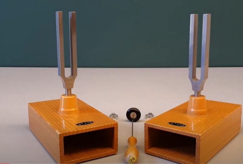
For the tuning fork experiment , students should have two identical wooden boxes with an open end. They need to have similar tuning forks placed at the center of the box. When the students strike tuning fork, the resonance in the box results in the amplification of the sound. If there is a box only a few centimeters away from the open end of another box, a strike on one of the tuning forks initiates a sympathetic vibration in the other one.
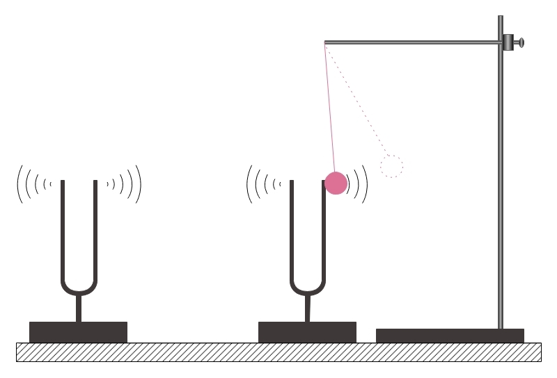
It can be observed by lightly touching the sound of the fork with a ping-pong ball tied to a thread. When the fork vibrates, the ball bounces back and forth. Students can carry out the demonstration with the help of a lump of clay. It can show a reduction in the frequency of the fork, and when struck, generate clear beats.
Process: This experiment aims to show how sound increased with resonance. For the study, the boxes must be there to face each other's open ends. Then, the ping-pong ball stand is fixed in such a way that it softly touches the fork. If one of the tuning forks is stuck during the experiment, students observe a back-front movement of the ping-pong ball. When a lump of clay is there at the fork's tine, each striking fork creates beats that the students can hear. With the increase in the size of clay, the frequency decreases.
3. How to Draw Tuning Fork Resonance Diagram
There are two ways to draw the tuning fork resonance experiment diagram, one is by hand, and another is use the easiest online tool.
3.1 How to Draw Tuning Fork Resonance from Sketch
The students can create the scientific diagram of the tuning fork resonance experiment by hand. For that, they need to follow these instructions:
Step 1: First, the students need to draw two hollow boxes which have their openings facing each other.
Step 2: Then they need to place the tuning forks on the top of the boxes, which look like the English alphabet 'U' with a stand at their bottom.
Step 3: Beside one of the tuning forks, there should be a stand with a ping-pong ball attached to a thread.
Step 4: The ball should touch the tuning fork, and there should be a symbol of sound drawn near both the tuning forks.
3.2 How to Draw Tuning Fork Resonance Online
Creating the diagram of scientific experiments by hand can be challenging. The students must use the EdrawMax Online tool. It can save time and help in drawing a high-quality image of a tuning fork resonance experiment. Here are a few simple steps which they can follow:
Step 1: EdrawMax Online is a user-friendly tool. Any inexperienced user can work on it comfortably. To start with their diagram, they need to go to the EdrawMax Online tool and open New . Next, they need to click on Science and Education option. Or the Science Illustration section.

Step 2: The tool is immensely beneficial for creating different educational pictures, which the students can use for their studies and projects. They can find the template in the Mechanics . You can select the diagram they need from here.

Step 3: EdrawMax Online tool allows the users to edit the images to suit their requirements. After selecting the template, you can easily modify it as per their choice.
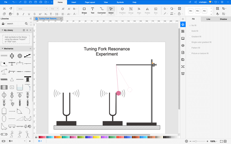
Step 4: Finally, when the student completes their diagram, they can easily save it and Export in different formats for using it in their projects. It supports to export in mulitple formats, such as Microsoft Office, Graphics, PDF, and more. Or, you even can present it in front of others by using the Presentation Mode .

4. The Resonance Examples
Resonance has everyday usage. In daily life, starting from the microwave oven to the gigantic bridge, resonance has something to do with them. Moreover, many famous musicians are grateful for this phenomenon called resonance because many musical instruments work on this principle. Here are a few examples of resonance in everyday life
- Guitar: When the guitarist strikes the guitar strings, there is a vibration. It eventually gets passed on to the hollow wooden box. Thus, creating resonance, and the sound gets amplified.
- Radio: There should be a change in the natural frequency of the receiver to listen to a particular radio station. When the natural frequency of the receiver matches the radio station's transmission frequency, there is a transfer of energy that allows a person to listen to a particular channel.
- Bridge: The soldiers are often not allowed to march on the bridge. The vibration created by the march on the bridge added to the bridge’s natural frequency can cause it to fall apart. In the case of the Tacoma bridge, the frequency of air matched that of the bridge.
- Pendulum: The pendulum shows a regular sidewise movement in a regular time interval. When the pendulum reaches its natural frequency of oscillation without even a push, it maintains the same amplitude.
- Microwave oven: There is a particular wavelength and frequency of the microwave oven radiation while the molecules of water and fat have their resonance. They absorb the wavelength at a specific frequency and vibrate. It causes the food to heat up for cooking.
5. Conclusion
During an experiment of mechanics or physics, it is best to use scientific diagrams. For these subjects, experiments largely depend on the process. A correct setup and procedure can give an accurate result. Therefore, to learn about the tuning fork resonance experiment , the students should use scientific diagrams. However, it may be challenging to create a high-quality tuning fork resonance diagram by hand. The students and researchers must use the EdrawMax Online tool.
In conclusion, EdrawMax Online is a quick-start diagramming tool, which is easier to make artery and vein diagram and any 280 types of diagrams. Also, it contains substantial built-in templates that you can use for free, or share your science diagrams with others in our template community .
Related Articles

Thank you for visiting nature.com. You are using a browser version with limited support for CSS. To obtain the best experience, we recommend you use a more up to date browser (or turn off compatibility mode in Internet Explorer). In the meantime, to ensure continued support, we are displaying the site without styles and JavaScript.
- View all journals
- Explore content
- About the journal
- Publish with us
- Sign up for alerts
- Open access
- Published: 19 September 2024
Demonstration of shape analysis of neutron resonance transmission spectrum measured with a laser-driven neutron source
- Mitsuo Koizumi 1 ,
- Fumiaki Ito 1 nAff4 ,
- Jaehong Lee 1 ,
- Kota Hironaka 1 ,
- Tohn Takahashi 1 ,
- Satoshi Suzuki 1 ,
- Yasunobu Arikawa 2 ,
- Yuki Abe 2 ,
- Zechen Lan 2 , 3 ,
- Tianyun Wei 2 ,
- Takato Mori 2 ,
- Takehito Hayakawa 2 , 3 &
- Akifumi Yogo 2
Scientific Reports volume 14 , Article number: 21916 ( 2024 ) Cite this article
1 Altmetric
Metrics details
- Applied optics
- Applied physics
- Nuclear energy
- Nuclear physics
- Optical techniques
- Techniques and instrumentation
Laser-driven neutron sources (LDNSs) can generate strong short-pulse neutron beams, which are valuable for scientific studies and engineering applications. Neutron resonance transmission analysis (NRTA) is a nondestructive technique used for determining the areal density of each nuclide in a material sample using pulsed thermal and epithermal neutrons. Herein, we report the first successful NRTA performed using an LDNS driven by the Laser for Fast Ignition Experiment at the Institute of Laser Engineering, Osaka University. The key challenge was achieving a well-resolved resonance transmission spectrum for material analysis using an LDNS with a limited number of laser shots in the presence of strong background noise. We addressed this by employing a time-gated \(^{6}{\textrm{Li}}\) -glass scintillation neutron detector to measure the transmission spectra, reducing the impact of electromagnetic noise and neutron and gamma-ray flashes. Output waveforms were recorded for each laser shot and analyzed offline using a counting method. This approach yielded a spectrum with distinct resonances, which were attributed to \(^{115}\,{\textrm{In}}\) and \(^{109}{\textrm{Ag}}\) , as confirmed through neutron transmission simulation. The spectrum was analyzed using the least-square nuclear-resonance fitting program, REFIT, demonstrating the possibility of using an LDNS for nondestructive areal-density material characterization.
Demonstration of non-destructive and isotope-sensitive material analysis using a short-pulsed laser-driven epi-thermal neutron source
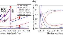
A new thermography using inelastic scattering analysis of wavelength-resolved neutron transmission imaging
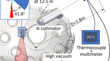
Single-shot laser-driven neutron resonance spectroscopy for temperature profiling
Introduction.
The continuous advancement of high-power laser technologies opens new possibilities for ion acceleration and applications using accelerated ions 1 , 2 , 3 . When a laser beam impacts a thin target, it accelerates ions through short-duration laser–plasma interaction near the target. The ion acceleration is determined by the characteristics of the target and laser, such as type and thickness of the target material, laser wavelength, polarization, and time and space density distributions 1 , 4 , 5 . The mechanisms are categorized into regimes, such as target normal-sheath acceleration (TNSA) 6 , 7 , radiation pressure acceleration (RPA) 8 , breakout afterburner (BOA) 9 , and collisionless shock acceleration (CSA) 4 . The maximum energy of the accelerated ions can reach tens of MeV, and even exceed 150 MeV 10 .
Utilizing laser-accelerated ions, laser-driven neutron sources (LDNSs) attract interest as short-pulse high-intensity neutron generators. These systems generate neutrons via reactions between the accelerated ions and a neutron converter (secondary target) near the laser target 3 , 4 , 5 , 11 . The neutrons have a broad energy distribution, with the maximum energy potentially as high as that of the accelerated ions. Furthermore, the size of the neutron converter (neutron source) can be reduced to only a few centimeters, because the laser-driven ion generation occurs in a micrometer-scale region at the target, and the neutron converter is placed near the target. The neutron generation duration is short, aligned with a laser pulse duration of less than a nanosecond, significantly shorter than that of accelerator-driven pulsed-neutron facilities 12 , 13 , 14 . Except for fast-neutron applications, a moderation system is crucial for extracting neutrons with the desired energy, i.e., epithermal 15 , thermal 16 , and cold 17 . Radiography experiments and nuclear reaction observation have been performed using neutrons of various energies 3 , 18 , 19 , 20 , 21 , 22 .
Neutron resonance analysis is a nondestructive assay (NDA) method using pulsed thermal-epithermal neutron beams 23 , 24 , 25 . This technique relies on neutron time-of-flight (n-TOF). In neutron resonance transmission analysis (NRTA), a sample is positioned between the neutron source and the detector. The neutron flight times from the source to the detector are measured at a known distance, and the neutron energies are deduced. The resulting n-TOF transmission spectrum shows nuclide-specific resonance patterns due to nuclear reactions, such as scattering, capture, and fission. The areal densities of each nuclide are determined by analyzing the spectrum. High-resolving power in NRTA is achieved through a long flight path length, a short neutron pulse duration, or both 23 , 24 , 26 . LDNSs could suit the NRTA measurements because of their ability to generate short neutron pulses. This capability allows for a compact n-TOF system, making NRTA more efficient.
The measurement challenges are high-flux neutron detection in high-background environments, such as the electromagnetic (EM) noise and strong gamma-ray and neutron flashes at the laser shot. The nuclear reactions of neutrons and ions with surrounding materials induce gamma-ray emissions, which can saturate and destabilize the detector output signal and cause after-pulsing in photomultiplier tubes (PMTs) 27 . A PMT with gated voltage can reduce the impact of the flash signals 28 , 29 , 30 . Neutron resonance transmission experiments using LDNSs have been reported 31 , 32 , 33 , 34 . Resonant fast-neutron absorption at 3 MeV in a graphite sample was observed using a plastic scintillator connected to a silicon photomultiplier in a noise shield via an optical fiber 31 . Drops in neutron counts at the neutron resonance energies in a spectrum were observed from 18 laser shots using a borated microchannel plate (MCP) detector, which has low sensitivity to gamma rays 32 . The changes in neutron transmission at the neutron resonance energies were observed by monitoring the output level of a time-gated \(^6\) Li-glass scintillation neutron detector with a single laser shot 33 . Furthermore, temperature monitoring was demonstrated by observing the broadening of resonance using single-shot neutron pulse beams 34 . These studies suggested the potential of neutron transmission resonance measurements for determining the nuclear areal densities in a sample.
Herein, we report the first successful nuclear areal-density analysis using neutron transmission spectra obtained by employing an LDNS. These spectra were measured using a time-gated \(^6\) Li-glass scintillation neutron detector to reduce the impact of EM noise as well as neutron and gamma-ray flashes. The output waveforms from the detector were recorded for each laser shot. The data were analyzed offline using a counting method to reduce uncertainty in the detector response. A spectrum with distinct resonances was obtained from merely three laser shots. The assignment of the resonances attributed to \(^{115}\,\hbox {In}\) and \(^{109}\,\hbox {Ag}\) in the sample was confirmed via simulations, which also ruled out spurious detector responses. The obtained spectrum was analyzed using the least-square nuclear resonance fitting program called REFIT 35 , demonstrating the feasibility of nondestructive areal-density measurement of nuclides in a sample using an LDNS.
Experimental setup
An experiment was performed using an LDNS 33 with the Laser for Fast Ignition Experiment (LFEX) 36 at the Institute of Laser Engineering (ILE), Osaka University. Figure 1 shows the experimental setup. The LDNS was placed at the target location of the LFEX, at the center of a stainless steel (Steel Use Stainless: SUS) vacuum chamber with a radius of 0.8 m. The pulse width of the LFEX was approximately 1.5 ps at full width at half maximum (FWHM), and the total energy was approximately 1000 J. The laser intensity at the LDNS target was approximately \(10^{19}\, \hbox {W/cm}^{2}\) . The laser repetition rate was limited to three shots per day due to the required cooling after each laser shot.

A diagram of the neutron-resonance transmission measurement setup using a laser driven neutron source (LDNS): ( a ) overview of the experimental setup with the neutron flight path; the sample used in the experiment was a stack of 0.2-mm-thick indium ( \(^{nat}\hbox {In}\) ) and 0.8-mm-thick silver ( \(^{nat}{\textrm{Ag}}\) ) plates; ( b ) a drawing of the LDNS comprising the first target (CD film) for charged-particle acceleration, the second target (neutron converter) for neutron generation, and the polyethylene moderator for neutron deceleration.
The LDNS comprised a target, a neutron converter, and a moderator (Figure 1 (b)). The target was a 5- \(\mu\) m-thick deuterated polystyrene (CD) foil, which was evaporated and replaced for each laser shot. The neutron converter comprised two beryllium (Be) rods, each 0.5 cm in diameter and 1 cm in height. The moderator was a 4-cm-thick truncated cone-shaped polyethylene block. When a laser pulse was shot on the CD target, deuterons and protons on the target surface were accelerated through the laser–plasma interaction. Neutrons were generated via the \(^{9}\) Be(d, n) and \(^{9}\) Be(p, n) reactions in the neutron converter, which were subsequently decelerated in the polyethylene moderator.
A neutron flight path was arranged at a horizontal angle of \({40}^\circ\) to the laser beam injection, and an Al window was set on the vacuum chamber. Collimators with a 5 cm \(\times\) 5 cm aperture comprising 20 \(\%\) borated polyethylene and Pb blocks (mostly sized 5 cm \(\times\) 10 cm \(\times\) 20 cm) were installed along the neutron flight path. A 1-cm-thick Pb plate was placed in the neutron beam path to reduce gamma-ray flashes from the laser plasma. At the end of the beam path, approximately 3.6 m from the moderator surface, a scintillation neutron detector was positioned, within a Pb-shielded space. The neutron detection system is detailed in the Methods section. The collimated neutron beams were directed solely onto the scintillator to prevent gamma rays and neutrons from directly hitting the PMT. The output waveforms from the neutron detector were recorded and analyzed as described in the Methods section.
Neutron transmission spectra
Figure 2 (a) shows the experimentally obtained n-TOF spectrum. In the measurement, a stack of 0.2-mm-thick indium ( \(^{nat}\,\hbox {In}\) ) and 0.8-mm-thick silver ( \(^{nat}\,\hbox {Ag}\) ) plates was used. The samples and their thicknesses were chosen to measure the resonance dips. The recorded shot-by-shot waveforms were analyzed offline to extract the n-TOF spectrum (see Methods). Three resultant spectra were averaged, including one measured on a separate day. The ordinate of the spectrum represents the averaged neutron counts in a 7- \(\mu\) s time bin, with a unit of counts per \(\mu\) s per laser shot. The statistical uncertainty in transmission within a 7- \(\mu\) s time bin obtained by three laser shots was approximately 5%. The energy resolution of the 7 \(\mu\) s time bin corresponds to 1%–7% for resonance energies from 0.5 eV to 25 eV, where the resonance broadening due to the moderator was approximately 1%, as evaluated via the simulation presented in the Methods section.

The n-TOF spectra through a stack of \(^{nat}\,\hbox {In}\) and \(^{nat}\,\hbox {Ag}\) plates: ( a ) the experimental and simulated n-TOF spectra; the ordinate represents the averaged neutron counts in a 7- \(\mu\) s time bin, with a unit of counts per \(\mu\) s per laser shot; the filled circles show the experimental data, the black line is to guide the eye, the solid and dashed blue lines show the simulation results with and without the sample, respectively; ( b ) the neutron transmission spectra; the experimental data points (closed circle) and a blue line connecting the data points obtained by the fitting program, REFIT, are presented; ( c ) the neutron transmission spectra calculated using nuclear data 38 ; the thickness of the In plate was varied while that of the Ag plate was kept 0.8 mm; the thicknesses of the In plate are indicated on the right-hand side of the panel; the transmission was the averaged value of the 7- \(\mu\) s time bin.
The resonance dips appeared at approximately 110 and 220 \(\mu\) s, where the resonances were attributed to \(^{109}\) Ag (5.19 eV, 2.32 \(\times\) \({10}^{4}\) b) and \(^{115}\,\hbox {In}\) (1.46 eV, 2.93 \(\times\) 10 \(^{4}\) b), respectively. The results correlated with the evaluated values of 114 \(\mu\) s and 215 \(\mu\) s using the nonrelativistic relation, TOF ( \(\mu\) s) = \(72.3 \times L/\sqrt{E}\) , where E is the neutron energy in eV, and L is the flight path length (3.6 m). The depth of the resonance dips is approximately half of the counts at the center of the dips, around 170 \(\mu\) s, whereas the neutron yield at the dips in the simulation is less than 10% (see Figure 5 (a) in the Methods section). The neutron-capture gamma-ray background from the neutron source can explain this discrepancy 39 .
Transmission spectra deduced from the simulated neutron flux presented in the Methods section are also plotted in Figure 2 (a). The gamma-ray background counts were approximated using a linear function passing through the bottom of the dips in the experiential spectrum, and the energy-dependent neutron detection efficiency of the \(^{6}\) Li-glass scintillation detector was calculated using the \(^6\) Li(n, t) cross-sections from Japanese Evaluated Nuclear Data Library (JENDL) 4.0 38 . The simulated neutron counts were scaled to match the depth of the experimental ones, evaluating approximately 10 \(^{11}\) neutron generation per shot at the LDNS neutron converter. As shown in Figure 2 (a), the simulated spectrum matched the experimental results, confirming the successful observation of resonances without any spurious contribution from neutron and gamma-ray flushes as well as EM noise.
Resonance shape analysis
The present experimental data was analyzed using a least-square fitting program, REFIT 35 . Figure 2 (b) shows measured neutron transmission data points (closed circle) and the fitting result (blue line). The experimental transmission was calculated by applying the background linear function, which was used for the adjustment of simulated spectra to the experimental data, and a linear function fitted for the simulated spectrum without the samples (see Figure 2 (a)). The error bars on the experimental data points were from experimental statistics. The uncertainty from the background subtraction and normalization were disregarded, assuming they were measured separately with good statistics. The fitting was performed for determining the areal densities of \(^{109}\) Ag and \(^{115}\) In, the flight length, and the initial delay of the n-TOF spectrum. As the contributions of \(^{107}\) Ag and \(^{113}\) In to the spectrum are comparably smaller than those of \(^{109}\) Ag and \(^{115}\) In, their areal densities were fixed to prevent unexpected effect on the fitting. Instead, their areal densities were manually adjusted to maintain consistent isotopic abundance. The fitting was repeated until the changes in the variables settled within their errors. Table 1 shows the resultant fitting values of REFIT. The flight length is consistent with our experimental setup. The initial delay correlate with the expected value of –3.5 \(\mu\) s, due to the bunching of the spectrum for 7 \(\mu\) s. The resultant areal densities correlated within the errors with the areal densities: 2.25 \(\times\) 10 \(^{-3}\) at/b for \(^{109}\) Ag and 0.734 \(\times\) 10 \(^{-3}\) at/b for \(^{115}\) In. The uncertainty of the determined areal densities is approximately 25%. These results confirm the viability of an LDNS for neutron resonance transmission measurements, demonstrating areal density determination with minimal laser shots.
For real sample measurements, additional measurements are required, i.e., a measurement without a sample to determine neutron transmission, and a measurement with filtering plates to evaluate the gamma-ray background by resonantly blocking neutrons. In parallel, the neutron yield is measured using a neutron flux monitor placed elsewhere to normalize the obtained spectra. This method, NRTA, applies to an areal density measurement of nuclides with resonances in an appropriate energy range, typically from thermal to epithermal ranges, where characteristic resonances are observed. These nuclides are mostly heavy elements including nuclear materials.
Figure 2 (c) shows the neutron transmission spectra using different stacks of \(^{nat}\) In and \(^{nat}\) Ag plates calculated using nuclear data 38 . The transmission data points are averaged in 7- \(\mu\) s time bins, which is the same as the present n-TOF spectrum time resolution. As the thickness of the In plate increases, the resonance depth of 1.46 eV (29.3 \(\times 10^3\) b at peak) increases, saturates, and then broadens, showing the applicability of the employed system for measuring the \(^{nat}\) In areal density relevant to thicknesses from 0.01 mm to 2.0 mm. Resonance dips with comparably low cross-sections at 3.82 eV (0.952 \(\times 10^3\) b) and 9.07 eV (1.50 \(\times 10^3\) b) appear with sample thicknesses greater than 0.5 mm, and the resonances of \(^{109}\) Ag (5.19 eV) and \(^{115}\) In (3.82 eV) form superimposed dips.
The uncertainty of this system can be reduced by increasing the statistics. Acceptable neutron detection numbers in the same energy range can be increased by extending the flight path, which prolongs the arrival time of neutrons within a specific energy range. This allows the use of a more efficient detector with sufficient thickness and a larger detection area. Furthermore, detectors capable of managing high count rates, such as segmented detectors, are effective. In the future, the repetition rate of laser systems should increase. A diode laser excitation method will enhance the repetition rate and reduce the size of the laser system. Petawatt-class lasers with 1–10Hz repetition rates are being developed 40 , 41 , 42 , 43 , 44 . The advancement of laser power density should increase neutron flux per laser shot. The target must be replaced quickly to catch up with the laser frequency because a single strong laser shot evaporates it. Target systems using target arrays, liquid targets, and other approaches, are proposed and being actively developed 45 , 46 , 47 .
The neutron generation of this experiment was evaluated approximately 10 \(^{11}\) n/shot, whereas the references indicate 1.6 \(\times 10^{10}\) n/shot 32 and 2–3 \(\times 10^{11}\) n/shot 33 . The difference in the neutron yields resulted from the target–converter–moderator combination and laser power density. The neutron generation is equivalent to or more than those of the electron accelerator-driven n-TOF facilities, such as the Geel Electron Linear Accelerator (GELINA; 3.4 \(\times 10^{13}\) n/s, 800 Hz; equivalent to 4.3 \(\times 10^{10}\) n/shot) 12 and the Hokkaido University Neutron Source facility (HUNS; 1.6 \(\times 10^{12}\) n/s, 100 Hz; equivalent to 1.6 \(\times 10^{10}\) n/shot) 14 . The neutron generation yield with LDNSs can be increased with laser power. A relation proposed for neutron generation n (neutron/shot) using an LDNS is \(n \propto I^4\) , where I is the laser power density (W/cm 2 ) 33 .
An advantage of LDNSs is the compactness of the system due to the small size of the area of accelerated particle generation, neutron converter, and moderator, all placed nearby. For example, the neutron generator of the KURRI-LINAC facility, which uses accelerated-electron-induced bremsstrahlung gamma rays to generate neutrons by the ( \(\gamma\) , n) reaction, comprising a water-cooled tantalum neutron converter ( \(\phi 50\) mm \(\times 60\) mm) and a water tank for neutron moderation (an octagonal ring of 300 mm \(\times\) 300 mm \(\times\) 100 mm), where the neutron converter is placed in the center of the moderator 13 . An n-TOF measurement system with a small moderator reduces the bore radius of the neutron flight path, reducing the volume of the collimator and shielding materials required. The short-pulse width of neutron generation, facilitated by subnanosecond laser pulses, enhances the time resolution of the n-TOF system, allowing for a shorter flight path to achieve a specific energy resolution, thereby increasing neutron flux. Another notable feature of LDNSs is that optical devices are used to transport laser beams, which are likely easier to control and tune than the heavy electromagnetic devices used in accelerator-driven neutron sources.
In conclusion, this study demonstrated the observation of a neutron resonance transmission spectrum using neutron beams from an LDNS with only three laser shots. The resonance spectrum was successfully analyzed, yielding the sample areal densities. This highlights the potential of a new approach to neutron-based nondestructive material characterization using an LDNS with advanced laser and target systems.
Neutron beam measurement
Figure 3 (a) shows the neutron detector system schematically. A \(^{6}\) Li-glass scintillator (KG2, 50 mm \(\times\) 50 mm \(\times\) 1 mm) was used due to its short decay time (18–62 ns), where the density of KG2 was 2.42 g/cm \(^3\) and included 7.5 wt% of Li (95% \(^6\) Li enriched) 48 . The thickness was chosen to achieve a suitable detection efficiency of 30 \(\%\) –6 \(\%\) for the 0.5–15 eV neutrons and to ensure that the detector responded to high-flux neutrons. A fused quartz plate (SiO \(_{2}\) , 50 mm \(\times\) 50 mm \(\times\) 4 mm) was used for a light guide, whereas an acrylic light guide was avoided to minimize neutron scattering. The scintillator and the glass plates were surrounded by reflector foil (aluminized Mylar), stored in a 1.5-mm-thick Al cap, and mounted on a PMT (R5113-02, a discontinued product of Hamamatsu), with a high-voltage divider and a transistor-switched gating circuit (C1392-11 MOD (normally on type), a discontinued product of Hamamatsu) 28 , 29 . The PMT was a 51-mm-diameter head-on type with a silica glass window. The photocathode was bialkali with an effective area of 46 mm in diameter, and the response wavelength was 160–650 nm.

( a ) Schematic of the detector system. The neutron beams passed through a \(^6\) Li-glass scintillator (KG2, 1 mm thick) with a light guide (fused silica (SiO \(_2\) ), 4 mm thick). The components were stored in an Al cap and mounted on a photomultiplier tube (PMT) with a transistor-switched gating circuit (normally on type). Two high-voltage power supplies (HVPSs) were used for the detector system. The positive voltage was imposed on the cathode using the gating circuit to suppress photoelectron multiplication. The detector output waveform was recorded using an oscilloscope. The measurement timings were controlled using a logic synthesizer. ( b ) A part of the recorded waveform from –20 to 200 \(\mu\) s. The PMT output saturation by gamma-ray and neutron flashes was avoided using the gated circuit of the detector at the beginning, and the elimination lasted at approximately 42 \(\mu\) s. The signals appeared after the gate period. The baseline shift and destabilization of the signal gradually recovered. ( c ) A part of an expanded waveform of Fig. 3 ( b ) between 160 and 164 \(\mu\) s. The gray line is the raw waveform. The black line is a corrected waveform using moving averages. The negative pulse signals under the threshold given by the dashed line are counted.
The neutron detector system was driven by two high-voltage power supply systems (HVPS) (3002D, Canberra): one for photoelectron multiplication and the other for the gating circuit. The photoelectron multiplication voltage imposed on the PMT was reduced from the recommendation of –2.0 kV to –1.4 kV to enhance the over-current resistance caused by a rush of signals. A logic synthesizer (Broad3, Bee Beans Technologies) triggered the gating circuit of the detector and a 12-bit analog-to-digital converter oscilloscope (MSO 58, Tektronix). The start timing signal was sent from the LFEX facility. The gating circuit of the PMT blocked the response of the detector for a fixed time of approximately 10 \(\mu\) s. To avoid the saturation of the PMT output by the laser-induced flashes, the gate period was extended by repeating 10 consecutive gate signals every 4 \(\mu\) s. The detector output waveforms were recorded using the oscilloscope in a 0.5 V full-scale range for 1 ms, with a sampling rate of 1.25 GS/s. The achieved waveform was analyzed offline.
Figure 3 (b) shows a part of the measured waveform. The gate circuit of the detector eliminated the output signal at the beginning of the waveform. The strong spike signal at the laser shot ( \(t = 0\) \(\, \mu\) s) was from the EM noise, and the other spikes were attributed to the gating circuit. The detector signal appears after the gate period ( \(t > 42\) \(\, \mu\) s), and the shifted and destabilized baseline gradually recovered. The gray line in Figure 3 (c) shows an expanded part of Figure 3 (b). The output amplitudes of the neutron signals were not significantly larger than the electronic noises. Therefore, in this analysis, the electronic noises were reduced using two moving averages, where the averaged values were assigned to the center of each averaging window. One moving average was calculated over 6.4 \(\mu\) s to remove the long-range baseline shift. The other was a short-range moving average over nine data points in 7.2 ns to remove the sharp electric noise.
The neutron detection timing was determined using the leading-edge method instead of constant fraction discrimination because the leading-edge method is straightforward, and the detector signal was fast enough to achieve the required time resolution (microsecond range). Negative pulse signals exceeding a threshold level were counted, indicated by the dashed line in Figure 3 (c). The threshold level was set to make the resonance dips appear clearly in the observed spectrum, resulting in a high threshold level to minimize noise signal counting. A 40-ns software dead time after pulse detection was imposed to avoid double counting the pulse signals. This time range was sufficient to cover the tip of the peak of a neutron pulse signal, where a decay time of 90%–10% of the signal of KG2 is 93 ns 48 . Figure 2 (a) shows the resultant spectrum.
The neutron counting rate increases as the flight time decreases (i.e., as the neutron energy increases), as shown in the spectra of Figure 2 (a). This makes it challenging to measure high-energy neutron resonances due to the high event rate. The maximum neutron energy available with this system was approximately 25 eV (corresponding to \(\sim\) 50 \(\mu\) s), where the baseline level of the PMT output stabilized (Figure 3 (b)). One can measure neutron resonances at higher energies by increasing the flight path length to delay the measurement timing or replacing the neutron detector with one that is noise-resistant and capable of managing high count rates.
Simulation of the neutron resonance transmission experiment
To evaluate the performance of the present n-TOF system, a neutron transmission simulation was conducted using the Particle and Heavy Ion Transport code System (PHITS) Ver. 3.12 37 , with the Japanese Evaluated Nuclear Data Library (JENDL)-4.0 38 . The neutron energy distribution from the neutron converter (Figure 4 (a)) employed in the simulation was based on the results of a simulation reported in a previous study 49 , wherein neutrons were generated from a 5-mm Be neutron converter placed 2 mm apart from a DC target via interaction with incident protons and deuterons with experimentally obtained energy distributions 49 . The uniformly generated neutrons from the neutron converter were slowed down in the moderator, and then flew to the 5 cm \(\times\) 5 cm detector (Figure 1 ). This simulation included a stainless-steel vacuum chamber, Al window, Pb plate, and parts of the polyethylene (20 \(\%\) borated) and Pb collimators (Figure 1 ). Figures 4 (b) and (c) show the neutron energy distribution at the detector, while Figure 4 (c) shows the thermal to epithermal energy region. Figure 5 (a) shows the resultant n-TOF spectra with and without the sample, comprising a stack of 0.2-mm-thick indium ( \(^{nat}\) In) and 0.8-mm-thick silver ( \(^{nat}\) Ag) plates. Strong resonance dips of 1.46 eV of \(^{115}\) In and 5.19 eV of \(^{109}\) Ag were observed, where the transmissions fell below 10%. Approximately 10 \(^{-10}\) neutrons per source per \(\mu\) s were expected in a 25 cm \(^2\) neutron detector area.

The energy distributions of neutrons. ( a ) The incident neutron energy distribution at the entrance of the moderator of the LDNS used for this PHITS simulation study. This distribution was obtained from the results of a simulation described in ref. 49 , which involved a 5-mm thick Be neutron converter positioned 2 mm from a DC target, and incident protons and deuterons with energy spectra derived from an experiment conducted at LFEX. Approximately, 50% of neutrons are less than 3 MeV and 80% are less than 5 MeV. ( b ) The neutron energy distribution from 0 MeV to 12 MeV at the detector position. ( c ) The neutron energy distribution from 0 eV to 25 eV at the detector position.
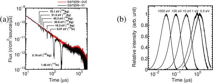
Results of simulation of the Particle and Heavy Ion Transport code System (PHITS) 3.12 37 . ( a ) Neutron time -of-flight (n-TOF) spectra simulated with 3 \(\times\) 10 \(^{11}\) neutron generations. The red line indicates the neutron flux (sample-out). The black line indicates the neutron flux through a sample (sample-in), i.e., the transmission n-TOF spectrum. All the fluxes are normalized by one neutron generation at the source. Assignments of strong resonances are given. ( b ) Time distribution of neutrons with same kinetic energies coming out from the moderator surface after the laser shot. The peak heights were normalized. The full width at half maximums (FWHMs) of the time distribution of 0.5, 1, 10, 100, and 1000 eV are 1.8, 1.3, 0.39, 0.13, and 0.042 \(\mu\) s, respectively.
The energy resolution in a neutron resonance-spectrum measurement can be expressed by \(\Delta E_n/E_n = 2\Delta t/t = 2 \Delta t \sqrt{2E/m_n }/L\) , where \(E_n\) and \(\Delta E_n\) are the neutron kinetic energy and its width, respectively, t and \(\Delta t\) are the neutron flight time and its broadening, respectively, \(m_n\) is the neutron mass, and L is the flight path length. The time broadening ( \(\Delta t\) ) is determined by the duration of neutron generation (less than nanosecond for LDNSs), the neutron moderation, and the geometrical flight path differences ( \(\sim\) \(\Delta L/v\) , where \(\Delta L\) and v are the flight path difference and the neutron velocity, respectively). Neutron moderation contributes the most to the time broadening of LDNS.
Figure 5 (b) shows the simulated time structure of neutrons with identical kinetic energies at the front surface of the moderator (Figure 1 ). The width of the time distribution narrows as the neutron energy increases. The FWHMs of the time distribution of 0.5, 1, 10, 100, and 1000 eV are 1.8, 1.3, 0.39, 0.13, and 0.042 \(\mu\) s, respectively. The broadenings were used to evaluate the energy resolutions at a flight path of 3.6 m to be approximately 1 \(\%\) for neutron energies from 0.5 eV to 1 keV. This result correlates with the experimental value of \(\Delta E _{n} /E_{n} = 2.3\%\) for a 5.2-eV resonance dip measured with a 1.8-m flight path using the same LDNS 33 . This resolution, achieved using a short-pulse laser and a compact neutron moderator, is sufficient for resolving resonances in a spectrum for general purposes.
Data availability
The data that support the findings of this study are available from the corresponding author, M. K., upon reasonable request.
Macchi, A., Borghesi, M. & Passoni, M. Ion acceleration by superintense laser plasma interaction. Rev. Mod. Phys. 85 , 751 (2013).
Article ADS CAS Google Scholar
Daido, H., Nishiuchi, M. & Pirozhkov, A. S. Review of laser-driven ion sources and their applications. Rep. Prog. Phys. 75 , 056401 (2012).
Article ADS PubMed Google Scholar
Roth, M. et al. Bright laser-driven neutron source based on the relativistic transparency of solids. Phys. Rev. Lett. 110 , 044802 (2013).
Article ADS CAS PubMed Google Scholar
Huang, C.-K. et al. High-yield and high-angular-fluence neutron generation from deuterons accelerated by laser-driven collisionless shock. Appl. Phys. Lett. 120 , 024102. https://doi.org/10.1063/5.0075960 (2022).
Horný, V. et al. High-flux neutron generation by laser-accelerated ions from single- and double-layer targets. Scientific Reports 12 , 19767 (2022).
Article ADS PubMed PubMed Central Google Scholar
Hatchett, S. P. et al. Electron, photon, and ion beams from the relativistic interaction of petawatt laser pulses with solid targets. Phys. Plasmas 7 , 2076–2082 (2000).
Wilks, S. C. et al. Energetic proton generation in ultra-intense laser-solid interactions. Phys. Plasmas 8 , 542–549 (2001).
Robinson, A. P. L., Zepf, M., Kar, S., Evans, R. G. & Bellei, C. Radiation pressure acceleration of thin foils with circularly polarized laser pulses. New Journal of Physics 10 , 013021 (2008).
Article ADS Google Scholar
Yin, L. et al. Three-dimensional dynamics of breakout afterburner ion acceleration using high-contrast short-pulse laser and nanoscale targets. Phys. Rev. Lett. 107 , 045003 (2011).
Ziegler, T. et al. Laser-driven high-energy proton beams from cascaded acceleration regimes. Nature Physics 20 , 1211–1216 (2024).
Article CAS Google Scholar
Alejo, A. et al. Recent advances in laser-driven neutron sources. Il Nuovo Cimento C 38 , 188 (2016).
ADS Google Scholar
Ene, D. et al. Global characterisation of the GELINA facility for high-resolution neutron time-of-flight measurements by Monte Carlo simulations. Nucl. Instr. Meth. in Phys. Res. A 618 , 54–68 (2010).
Sano, T. et al. Analysis of energy resolution in the KURRI-LINAC pulsed neutron facility, in Proc. ND2016 03031 (Bruges, Belgium, 2017).
Anderson, I. S. et al. Research opportunities with compact accelerator-driven neutron sources. Phys. Rep. 654 , 1–58 (2016).
Mirfayzi, S. R. et al. Experimental demonstration of a compact epithermal neutron source based on a high power laser. Appl. Phys. Lett. 111 , 044101 (2017).
Mirfayzi, S. R. et al. A miniature thermal neutron source using high power lasers. Appl. Phys. Lett. 116 , 174102 (2020).
Mirfayzi, S. R. et al. Proof-of-principle experiment for laser-driven cold neutron source. Phys. Rep. 10 , 20157 (2020).
CAS Google Scholar
Yogo, A. et al. Single shot radiography by a bright source of laser-driven thermal neutrons and x-rays. Appl. Phys. Express 14 , 106001 (2021).
Wei, T. et al. Non-destructive inspection of water or high-pressure hydrogen gas in metal pipes by the flash of neutrons and x rays generated by laser. AIP Advances 12 , 045220 (2022).
Lan, Z. & Yogo, A. Exploring nuclear photonics with a laser driven neutron source. Plasma Phys. Control. Fusion 64 , 024001 (2022).
Mori, T. et al. Thermal neutron fluence measurement using a cadmium differential method at the laser-driven neutron source. J. Phys. G: Nucl. Part. Phys. 49 , 065103 (2022).
Mori, T. et al. Direct evaluation of high neutron density environment using (n, 2n) reaction induced by laser-driven neutron source. Phys. Rev. C 104 , 015808 (2021).
Postma H. & Schillebeeckx, P. Neutron resonance capture and transmission analysis, Meyers RA (ed) Encyclopedia of analytical chemistry , Wiley, New York, p. 1-22 (2009).
Schillebeeckx, P., Becker, B., Harada, H. & Kopecky, S. Neutron spectroscopy for the characterisation of materials and objects, European Commission Joint Research Centre, Geel, Belgium, Rep. EUR 26848-EN (2014).
Hironaka, K. et al. Neutron resonance fission neutron analysis for nondestructive fissile material assay. Nucl. Inst. Meth. in Phys. Res. A 1054 , 168467 (2023).
Tsuchiya, H., Kitatani, F., Maeda, M., Toh, Y. & Kureta, M. Development of neutron resonance transmission analysis as a non-destructive assay technique for nuclear nonproliferation. Plasma Fusion Res. 13 , 2406004 (2018).
Hamamatsu Photonics KK, Shizuoka, Japan, Afterpulsing, PHOTOMULTIPLIER TUBES: Basics and Applications , 3rd ed., Sec. 4.3.8, p. 77 (2007).
Hamamatsu Photonics KK, Shizuoka, Japan, Gating circuit, PHOTOMULTIPLIER TUBES: Basics and Applications , 3rd ed., Sec. 5.1.9, p. 97 (2007).
Hara, K. Y., Harada, H., Toh, Y. & Hori, J. γ-Flash suppression using a gated photomultiplier assembled with an LaBr 3 (Ce) detector to measure fast neutron capture reactions. Nucl. Instr. Meth. Phys. Res. A 723 , 121–127 (2013).
Abe, Y. et al. A multichannel gated neutron detector with reduced afterpulse for low-yield neutron measurements in intense hard X-ray backgrounds. Rev. Sci. Instrum. 89 , 10I114 (2018).
Article CAS PubMed Google Scholar
Kishon, I. et al. Laser based neutron spectroscopy. Nuclear Inst. Methods Phys. Res. A 932 , 27–30 (2019).
Zimmer, M. et al. Demonstration of non-destructive and isotope-sensitive material analysis using a short-pulsed laser-driven epi-thermal neutron source. Nat. Commun. 13 , 1173 (2022).
Article ADS CAS PubMed PubMed Central Google Scholar
Yogo, A. et al. Laser-driven neutron generation realizing single-shot resonance spectroscopy. Phys. Rev. X 13 , 011011 (2023).
Lan, Z. et al. Single-shot laser-driven neutron resonance spectroscopy for temperature profiling. Nat. Comm. 15 , 5365. https://doi.org/10.1038/s41467-024-49142-y (2024).
Moxon, M.C., Ware, T.C. & Dean, C.J. REFIT-2009 A least-square fitting program for resonance analysis of neutron transmission, capture, fission and ccattering data; User’s guide for REFIT-2009-10 (2010). report UKNSF(2010)P243 UK Nuclear Science Forum.
Miyanaga, N. et al. 10-kJ PW laser for the FIREX-I program. J. Phys. IV France 133 , 81–87 (2006).
Sato, T. et al. Features of Particle and Heavy Ion Transport code System (PHITS) version 3.02. J. Nucl. Sci. Technol. 55 , 684–690 (2018).
Shibata, K. et al. JENDL-4.0: A new library for nuclear science and engineering. J. Nucl. Sci. Technol. 48 , 1–30 (2011).
Lee, J. et al. Relationship between neutron moderator and time-dependent background for neutron time-of-flight measurement. J. Nucl. Sci. Technol. https://doi.org/10.1080/00223131.2023.2224330 (2023).
Article Google Scholar
Haefner, C. L. et al. High average power, diode pumped petawatt laser systems: a new generation of lasers enabling precision science and commercial applications, in Proc. SPIE 024102 (Prague, Czech, 2017).
Danson, C. N. et al. Petawatt and exawatt class lasers worldwide. High Power Laser Sci. Eng. 7 , 1–54 (2019).
Lureau, F. et al. High-energy hybrid femtosecond laser system demonstrating 2 × 10 PW capability. High Power Laser Science and Engineering 8 , e43 (2020).
Pathak, V. B. et al. Strong field physics pursued with petawatt lasers. AAPPS Bulletin 31 , 4 (2021).
Clark, E. L. et al. High-intensity laser-driven secondary radiation sources using the ZEUS 45 TW laser system at the Institute of Plasma Physics and Lasers of the Hellenic Mediterranean University Research Centre. High Power Laser Sci. 9 , e53 (2021).
Prencipe, I. et al. Targets for high repetition rate laser facilities: needs, challenges and perspectives. High Power Laser Sci. 5 , e17 (2017).
Poole, P. L. et al. Moderate repetition rate ultra-intense laser targets and optics using variable thickness liquid crystal films. Appl. Phys. Lett. 109 , 151109 (2016).
Noaman-ul-Haq, M. et al. Statistical analysis of laser driven protons using a high-repetition-rate tape drive target system Phys. Rev. Accel. Beams 20 , 041301 (2017).
Scintacor, https://scintacor.com/products/6-lithium-glass
Nishimura, H., Yasunobu, A., Yuki, A. & Akifumi, Y. Development of laser-driven neutron source and relevant diagnostics. Plasma Fision Res. 95 , 3–10 (2019) ( in Japanese ).
Download references
Acknowledgements
This work was implemented under the subsidy for “promotion of strengthening nuclear security and the like” of the Ministry of Education, Culture, Sports, Science, and Technology-Japan. The simulation work was conducted using the supercomputer HPE SGI8600 in the Japan Atomic Energy Agency.
Author information
Fumiaki Ito
Present address: High Energy Accelerator Research Organization (KEK), 1-1 Oho, Tsukuba-shi, 305-0801, Japan
Authors and Affiliations
Integrated Support Center for Nuclear Nonproliferation and Nuclear Security, Japan Atomic Energy Agency (JAEA), Tokai, Ibaraki, 319-1195, Japan
Mitsuo Koizumi, Fumiaki Ito, Jaehong Lee, Kota Hironaka, Tohn Takahashi & Satoshi Suzuki
Institute of Laser Engineering, Osaka University, Suita, Osaka, 565-0871, Japan
Yasunobu Arikawa, Yuki Abe, Zechen Lan, Tianyun Wei, Takato Mori, Takehito Hayakawa & Akifumi Yogo
Kansai Institute for Photon Science, National Institutes for Quantum and Radiological Science and Technology (QST), 8-1-7 Umemidai, Kizugawa-shi, Kyoto, 619-0215, Japan
Zechen Lan & Takehito Hayakawa
You can also search for this author in PubMed Google Scholar
Contributions
M.K., F.I., and J.L. conceived the experiment. M.K., F.I, J.L., K.H., T.T., and S.S. conducted the n-TOF experiment. M.K. supervised the experiments. Y.Ar., Y.Ab., Z.L., Z.L., T.W., T.M., T.H., A.Y., supported the experiments and provided neutrons using the LDNS. A.Y. supervised the operation of the LDNS. J.L. performed the simulation study. M.K. and F.I. analyzed the data. M.K., F.I., and J.L. wrote the manuscript. All authors reviewed the manuscript.
Corresponding author
Correspondence to Mitsuo Koizumi .
Additional information
Publisher’s note.
Springer Nature remains neutral with regard to jurisdictional claims in published maps and institutional affiliations.
Rights and permissions
Open Access This article is licensed under a Creative Commons Attribution-NonCommercial-NoDerivatives 4.0 International License, which permits any non-commercial use, sharing, distribution and reproduction in any medium or format, as long as you give appropriate credit to the original author(s) and the source, provide a link to the Creative Commons licence, and indicate if you modified the licensed material. You do not have permission under this licence to share adapted material derived from this article or parts of it. The images or other third party material in this article are included in the article’s Creative Commons licence, unless indicated otherwise in a credit line to the material. If material is not included in the article’s Creative Commons licence and your intended use is not permitted by statutory regulation or exceeds the permitted use, you will need to obtain permission directly from the copyright holder. To view a copy of this licence, visit http://creativecommons.org/licenses/by-nc-nd/4.0/ .
Reprints and permissions
About this article
Cite this article.
Koizumi, M., Ito, F., Lee, J. et al. Demonstration of shape analysis of neutron resonance transmission spectrum measured with a laser-driven neutron source. Sci Rep 14 , 21916 (2024). https://doi.org/10.1038/s41598-024-72836-8
Download citation
Received : 08 July 2024
Accepted : 11 September 2024
Published : 19 September 2024
DOI : https://doi.org/10.1038/s41598-024-72836-8
Share this article
Anyone you share the following link with will be able to read this content:
Sorry, a shareable link is not currently available for this article.
Provided by the Springer Nature SharedIt content-sharing initiative
By submitting a comment you agree to abide by our Terms and Community Guidelines . If you find something abusive or that does not comply with our terms or guidelines please flag it as inappropriate.
Quick links
- Explore articles by subject
- Guide to authors
- Editorial policies
Sign up for the Nature Briefing newsletter — what matters in science, free to your inbox daily.
A Method for Analyzing Data from 1- and 2-Dimensional Relaxation and Diffusion NMR Experiments by Determination of their Expectation Values and Standard Deviations
- Original Paper
- Open access
- Published: 20 September 2024
Cite this article
You have full access to this open access article

- Geir Humborstad Sørland 1 , 2 ,
- Henrik Walbye Anthonsen 2 &
- Sebastien Simon 1
A method for analyzing ill-posed multi-exponentially decaying data from 1- and 2-Dimensional experiments by Determination of the Expectation Values and their Standard Deviation has been developed. It combines a repeated use of the discrete Anahess approach for analyzing the dynamic data where a regrouping of the noise in the data is performed for each repetition. These resulting expectation values are used as initial and restricting values to produce a distribution using the Inverse Laplace transform, where position and volume of the distribution can then be reported with expectation value and standard deviation. The method is verified on synthetic data and tested on real data in both one and two dimensions.
A Robust Method for Analysing One and Two-Dimensional Dynamic NMR Data
Fitting a function to time-dependent ensemble averaged data
A data–driven approximation of the koopman operator: extending dynamic mode decomposition.
Avoid common mistakes on your manuscript.
1 Introduction
When analyzing relaxation and/or diffusion data acquired with various Nuclear Magnetic Resonance (NMR) techniques followed by the use of the Inverse Laplace transform (ILT) [ 1 , 2 , 3 , 4 , 5 , 6 , 7 , 8 , 9 ], the smoothing of the data is essential to produce distributions of relaxation times and/or diffusion coefficients. Even though there has been some work in non-uniform smoothing, the most common way to smooth the data is to apply a uniform smoothing on the data, i.e., a constant smoothing throughout the processing irrespective of the content of the data. Consequently, there will be a different broadening of the peaks depending on the peak position or fraction of the data in which the peak contributes to an attenuating signal [ 10 ]. Alternatively, discrete methods have been developed that fits the data to a limited number of components [ 11 , 12 , 13 ]. However, these methods suffer from the lack of information on the distributivity of the dataset.
Recently, a method was proposed that combines a discrete processing of the dataset together with the ILT, namely the Anahess distribution [ 10 ]. Here the number of components in the solution is limited to a minimum, which makes it possible to divide the solution into sub-groups that can be transformed and processed as superimposed datasets using the ILT with conditions set by the discrete solution. This approach has shown to reproduce synthetic distributions better than the ILT only. An extension of this method has now been developed and will be presented here. It aims at finding the expectation values for the fitted discrete components and the corresponding distribution. This should provide a measure allowing to evaluate the quality of the fit. In short, the procedure is as follows: after fitting the exponentially decaying data to a limited number of components according to the Bayesian Information stop Criterion (BIC) [ 14 ], a set of residual data or noise from the fit is produced. The residuals are then rearranged with respect to position. That is, the residuals or a group of such are interchanged in a random way so that new noise data is produced but with same expectation value and standard deviation as the original set of residuals. A new exponentially decaying data set can then be produced from the regrouped noise, and the fitted components are determined from the discrete fitting procedure. The new raw data set is then analyzed using the discrete Anahess, resulting in a new set of fitted components. If there is an impact of noise on the fitted components, the values will vary due to the different noise present. This procedure is repeated until enough data with rearranged residuals are produced to find an expectation value and a standard deviation for the fitted components.
In the following, we will recapture the combination of the discrete Anahess approach with ILT [ 10 ] and provide the method for determining the expectation values of the fitted components and distributions.
1.1 The Anahess Approach
In experimental data, the noise, ε, is superimposed on the exponentially decaying signal. Data arising from relaxation and/or diffusion NMR experiments can be described by a multi-exponential decaying signal S(t)
Here A(T k ) is the distribution of intensities to be fitted to the corresponding T k , which could be the corresponding relaxation time or the diffusion coefficient. The most common way to fit the equation above to experimental data is to use the Inverse Laplace Transform initially developed by Provencher [ 1 , 2 , 3 ]. Then, a predefined grid of a fixed number of points is defined, on which the solution ( A(T k ) ) has to be found. Another approach is to use a discrete method where the number of components is limited [ 11 ]. In this work, we apply the discrete Anahess approach, where the number of points (components) in the solution is minimized according to a Bayesian Information criterion and the points are allowed to move anywhere in the space of solutions. As in the ILT routine, a function involving the sum-squared relationship is to be minimized [ 11 , 15 ].
a 0 is a baseline offset which may be positive or negative. The data matrix R has the corresponding number of data points NX . The parameters a p , T p are thus characteristic properties of the component with index p out of all the NCO components. As SS re s will decrease with increasing number of fitted components, a stop criterion is needed. The Bayesian information criterion ( BIC ) [ 14 ] is such a criterion. Let n be the number of observed data points, let q be the number of free model parameters, and SS res be the sum of squared residuals. Then if the residuals are normally distributed, the BIC has the form:
In this equation, a good model fit gives a low first term while few model parameters give a low second term. When comparing a set of models, the model with the minimal BIC value is selected.
The discrete solution fits to a relatively small number of components that provide a satisfactorily fit of the raw data. This is due to the discreteness of the fitting routine when using the BIC as a stop criterion [ 14 ]. As most of data reflect continuous distributions of components, a method for probing the distributivity using the discrete Anahess results has been developed. This is done by applying the ILT where the results from the discrete Anahess fit are fed into the ILT as initial and restricting conditions [ 8 ]. Because of the limited number of components in the Anahess, one may group various regions in the solution and provide a superimposed fit of each group. Consequently, prior to the use of the ILT, the data are grouped and then transformed in t according to the following equation [ 10 ]
where t is the original observation time, T k is the result from the Anahess fit for component k , and t max is the longest observation time in the data set. With this approach, one may process data with the discrete Anahess method and probe the distributivity.
1.2 The Method for Determining the Expectation Value and its Standard Deviation
When the exponentially decaying data are fitted using the Discrete Anahess [ 11 , 15 ], a resulting set of residuals is produced. These residuals are the difference between the fitted and the original data. As the noise is the crucial part that affects the result of the fitting procedure, both using discrete and continuous fittings, we here propose to produce new raw data to be subjected for processing through redistributing the noise as follows: divide the residual noise into several compartments for both 1- and 2-dimensional noise, as shown in Fig. 1 . The compartments should be set so small that when moving them around in a random way such that the end product is a different noise dataset but with the same expectation value and standard deviation.
where j is a random number within the number of compartments. Using the fitted components and intensities from the Discrete Anahess method, one may then perform a Laplace Transform and produce new data sets where the noise is different due to the random interchanging of the compartments in the original noise data.
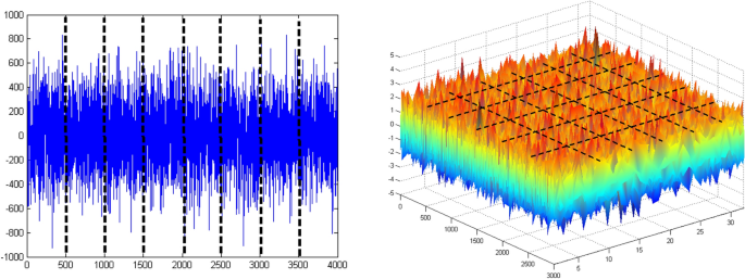
The synthetic Gaussian noise in one (left) and two (right) dimensions. The black dashed lines indicate the separation of the noise into different compartments (color figure online)
These datasets can then be analyzed using the discrete Anahess approach and a variation in the fitted intensities and new relaxation times and/or diffusion coefficients will be found. When this procedure has been repeated until the effect of the noise has been probed properly, it will result in an expectation value for the various components, and these values can then be used as initial and restricting values to probe the distributivity of the original dataset.
This approach assumes that there is no dependency of the noise as a function of observation time and/or applied gradient strength. That is, the discrete Anahess fit returns a Gaussian distribution of residuals that has an expectation value of 0 and the same insignificant skew [ 16 ]. Also, this method provides a way to check if the fit to the original raw data is a stable one. That is, the minimum BIC number is achieved at the same number of components and the variation in the expectation values is acceptable.
Once a set of expectation values are found for a data set, these values can be used as input when probing the distributivity using the ILT procedure. As the expectation values provide a good fit to the raw data, the ILT routine will probe the distributivity around these initial values, resulting in a distribution that is characteristic for the expectation values.
2 Experimental Section
A set of synthetic data sets in 1- and 2 dimensions were used to verify the proposed method for determining the expectation values [ 10 ]. The one-dimensional synthetic data set was produced from a distribution located on a grid of 200 T 2 values (0.0001–10 s) having three identical but separated peaks. After imposing a Laplace Transform to produce a decaying signal, synthetic Gaussian noise was added to mimic the experimental noise from an NMR experiment. The inter-echo spacing (2τ) was set to 0.4 ms and the data set contained 8000 echo points to mimic a Carr–Purcell–Meiboom–Gill (CPMG) decay. The two-dimensional synthetic data set was produced from distributions located on a 64 × 32 grid of T 1 ’s (0.001–10 s) and T 2 ’s (0.0001–10 s). As for the one-dimensional case, synthetic Gaussian noise was added, and the attenuation mimicked a combined Stimulated Echo—CPMG experiment [ 10 ].
In addition to the synthetic data sets, real NMR data sets were acquired from a sample of oat flakes. The NMR instrument applied was a 0.5 Tesla permanent magnet supplied by Advance Magnetic Resonance Ltd. [ 17 ] with the possibility of measuring samples of 18 mm in diameter. The one-dimensional experiment was a CPMG with inter-echo spacing of 0.2 ms acquiring 4000 echoes, and this was enough to secure the attenuating signal to reach the noise level. The Inversion Recovery (IR)-CPMG experiment was applied to produce two-dimensional data. After measuring on the oat flakes, the sample was dried at 105 C for 12 h and remeasured with the IR-CPMG experiment. The aim of this procedure is to identify the location of the moisture in the processed data prior to drying, and the oat flakes are chosen as sample because the T 2 signal from moisture and fat is found to partially overlap.
The discrete Anahess is the processing method that provides the fitted components used to find the expectation values and their standard deviation [ 18 ]. The application starts with fitting one component to a data set and calculate its BIC number, then proceeds to two components and finds a new BIC number. If the current BIC number is lower than the previous one, the application increases the number of components to be fitted by one, and a new BIC number is found. This procedure continues until the current BIC number is larger than the previous one. The best fit for the data set is then the NCO that produces the lowest BIC number. This is shown in Fig. 2 for NCO ∈ [ 3 , 7 ] using the data from the two-dimensional data set produced from oat flakes (Fig. 9 ), where the best fit is found at 6 components.

The fitted BIC number as a function of number of components
3 Results and Discussion
In the following, the results from processing of synthetic and real data in one and two dimensions are presented together with the expectation values and standard deviations achieved from the method proposed in Sect. 1.2
3.1 The Expectation Value for the One-Dimensional Synthetic Data Set
In Fig. 3 , the attenuation of the synthetic data set is shown together with the residuals from the discrete Anahess fit, and it resulted in a three-component fit for the lowest BIC number. In the upper right corner of the figure, the noise is plotted and fitted to a Gaussian distribution. The skew of the noise is found to be 0.003, which shows that the distribution is symmetrical around the expectation value 0. In other words, the discrete Anahess fit returns Gaussian noise as residuals, and one may produce new data sets by doing a random permutation of the noise compartments. For this experiment, the 8000 points of noise were divided into 80 compartments of 100 points each, and they were randomly permuted 10 times to provide 10 new raw data sets.
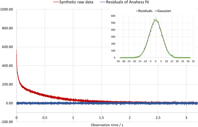
The attenuation of the three-exponential decaying signal (red curve) arising from a synthetic data distribution. The blue curve corresponds to the fitted residuals. In the upper right corner, the distribution of residuals is shown together with the fitted Gaussian curve (color figure online)
As the 10 datasets were processed, it turned out that the minimum BIC number was achieved at NCO = 3 for all data sets, and one could easily identify the components’ position throughout the series of fitted data. The results for the three components are found in Table 1 , while the synthetic and Anahess distribution are shown in Fig. 4 . The Anahess distribution is based on the average values found in Table 1 , and one may notice that the Anahess distribution is broadened due to the Gaussian noise that was added to the synthetic distribution when producing the original raw data. The areas of the synthetic peaks were 200 each, and the average T 2 values were 2.33, 35.80 and 568.57 ms. All average values in Table 1 were within the standard deviation except for the average intensity of the peak at longest T 2 , so the there is a good agreement between the fitted and key average values.

Comparison between the synthetic and the Anahess distributions produced from the average values for T 2 and initial intensities
What is evident from the data in Table 1 is an increasing relative standard variation as T 2 is reduced. For the peak at the shortest T 2 , the relative standard deviation is 4.2% for T 2 and 7.5% for the intensity, for the peak in the middle, the standard deviation is 1.3% for T 2 and 2.2% for the intensity, while for the peak at the longest T 2 , the standard deviation is 0.4% for T2 and 0.6% for the intensity. The reason for the decreasing uncertainty of the fitted values as T 2 increases is because the number of significant datapoints where the components contribute in the attenuating signal is increased as T 2 increases. For the component fitted to 2.39 ms, its signal has reduced to a fraction less than 0.01 at 24 ms. With a τ-value of 0.2 ms, this corresponds to 60 data points. Consequently, in the data that contains 8000 data points, the components with the shortest T 2 contribute only in 0.75% of the data. The component with T 2 of 35.38 contributes in 5.6% of the data while the component with T2 of 568.57 ms contributes in 88.9% of the data.
3.2 The Expectation Value for the One-Dimensional Real Data Set
In Fig. 5 , the attenuation of the data set arising from oat flakes is shown together with the residuals from the discrete Anahess fit resulting in three components with the lowest BIC number. The three components are one isolated component with T 2 ~ 0.6 ms and two components rather close to each other with T 2 ~ 50 and 180 ms respectively. In the upper right corner of the figure, the noise is plotted and fitted to a Gaussian distribution. The skew of the noise is found to be 0.03, which is the same skew as found from the synthetic Gaussian noise distribution, i.e., an insignificant skew. Thus, one may conclude that the distribution is symmetrical around the expectation value 0.001, and again the discrete Anahess fit returns Gaussian noise as residuals, and on may produce new data sets by doing a random permutation of the noise compartments. In this experiment, the 4000 points of noise were divided into 40 compartments of 100 points each, and they were randomly permuted 20 times to provide 20 new raw data sets.
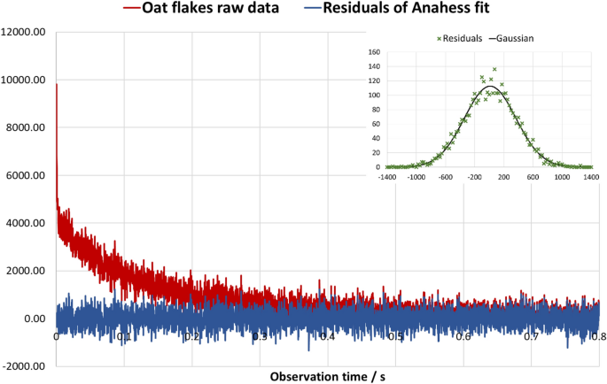
The attenuation of a decaying signal (red curve) arising from a CPMG experiment performed on oat flake. The blue curve corresponds to the fitted residuals. In the upper right corner, the distribution of residuals is shown together with the fitted Gaussian curve (color figure online)
As the 20 datasets were processed, it turned out that the minimum BIC number was achieved at NCO = 3 for all data sets, and one could easily identify the components’ position throughout the series of fitted data. The results for the three components are shown in Table 2 . The isolated component at the shortest T 2 is reported with a standard deviation of 9.1% in intensity and 13.3% in T 2 , while the component with intermediate T 2 (47.1 ms) is reported with higher standard deviations (14.2% in intensity and 21.4% in T 2 ). This does not fit the picture of an improved accuracy for the intermediate T 2 component as more datapoints are available. The reason for this is that the two components at longer T 2 do not correspond to two unique components as the varying noise significantly interferes with the fitted components. The solution is to group the two components together, as shown in Table 2 .The population weighted average of T 2 for the two components as a group is then calculated, and variation of this value in different fittings provides the expectation value and its standard deviation. Then the standard deviation for the group is down to 2.5% for the intensity and 3.2% for the T 2 . Thus, the improved accuracy of the expectation value for the group as compared to the individual components can be used as a criterion for the grouping of components. This knowledge can be applied when probing the distributivity of the data, and components 2 and 3 must be processed as a group. In Fig. 6 , the Anahess T 2 distribution is shown for the oat flakes, and it is based on the fit of the 20 data sets with different noise produced from the residuals of the fitting of the original data. Consequently, the intensity of the peaks and their average T 2 values (i.e., peak position) can be reported with a standard deviation given in Table 2 for component 1 (left peak) and component 2 + 3 (right peak). Alternatively, one may produce 20 data sets by running the experiment 20 times.
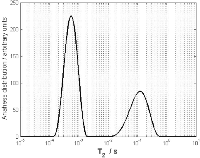
The Anahess T 2 distribution from oat flakes based on the average values for T 2 and initial intensities of the oat flakes components
3.3 The Expectation Value for the Two-Dimensional Synthetic Data Set
Figure 7 shows the results from the discrete Anahess fit of the two-dimensional synthetic data set, with a six-component fit for the lowest BIC number. In the right part of the figure, the noise is shown plotted and fitted to a Gaussian distribution. The skew of the noise was found to be −0.003, confirming that the distribution is symmetrical around the expectation value 0 and thus the discrete Anahess fit returns Gaussian noise as residuals. New data sets can then be produced by doing a random permutation of the noise compartments. For this example, the 3000 points of noise were divided into 30 compartments of 100 points each, and they were randomly permuted 10 times to provide 10 new raw data sets.

The discrete components from the Anahess fit to the left, the resulting residuals to the lower right and the distributions of residuals to the upper right
In Table 3 , the average results based on the 10 fitted data sets with varying noise are shown. As for the one-dimensional case on oat flakes, the components can be regrouped into component 4, components 1 + 2 and components 3 + 5 + 6. Without this grouping, the standard deviations for individual components at higher T 2 ’s are higher than the standard deviation for the component with shortest T 2 , while a lower standard deviation is to be expected due to more data points available for fitting the components of longer T 2 ’s. When grouping the components as shown in Table 3 , the standard deviation decreases as T 2 of the group increases. The Anahess distribution based on the average values of the components is shown in Fig. 8 , and it fits well to the synthetic distribution provided in [ 10 ].
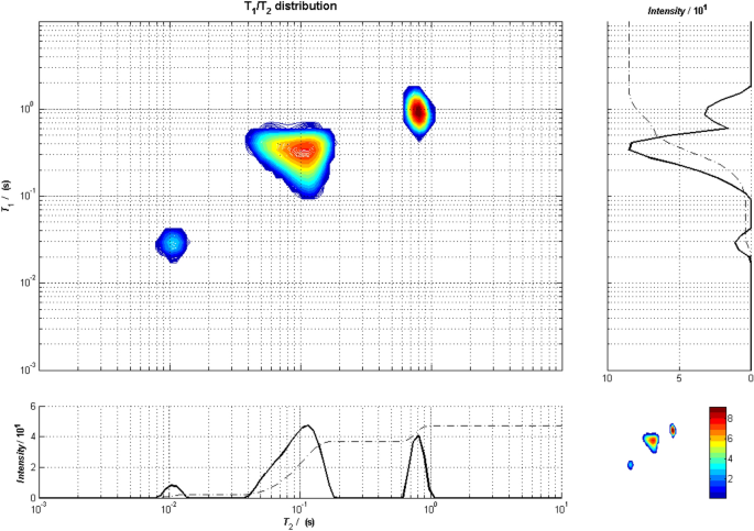
The Anahess T 1 –T 2 synthetic distribution based on the average values for T 1 , T 2 and initial intensities
3.4 The Expectation Value for the Two-Dimensional Real Data Set
Figure 9 shows the results from the discrete Anahess fit for a dataset recorded from a combined IR-CPMG experiment, and the lowest BIC number is found with a six-component fit. In the right part of the figure, the noise is shown plotted and fitted to a Gaussian distribution. The skew of the noise is found to be −0.002, confirming that the distribution is symmetrical around the expectation value 0, and thus the discrete Anahess fit returns Gaussian noise as residuals. New data sets can then be produced by doing a random permutation of the noise compartments. For this example, the 4000 points of noise were divided into 40 compartments of 100 points each, and they were randomly permuted 10 times to provide 10 new raw data sets.

When the 10 new datasets were processed using the discrete Anahess method, it was found that the lowest BIC number is 6 for 9 of the 10 datasets while for one data set, the lowest BIC number is found at NCO = 5. For this dataset, component 5 in Fig. 9 disappears and the neighboring components (6 and 3) are shifted slightly in position. This is a consequence of the Gaussian noise in combination with the fact that component 5 in Fig. 9 has an intensity of ~ 80 while the noise varies between ± 100. In short, in 10% of the processed data, we will fit to 5 components using Anahess while in 90% of the incidents, it will be reported 6 components. So based on the processed data, we can report the 6-component fit shown in Table 4 with a 90% probability. As for the synthetic two-dimensional dataset, we find that the standard deviation reduces as T 2 and T 1 increase if one regroups the components as component 4, components 1 + 2 + 3 and components 5 + 6. However, when producing the Anahess distribution as shown in Fig. 10 , it turns out that components 5 + 6 do not produce one peak as components 1 + 2 + 3 do. Thus, the probing of the distributivity indicates that component 5 should be separated from component 6.
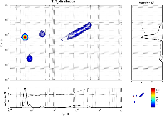
The Anahess T 1 –T 2 distribution from oat flakes based on the average values for T 1 , T 2 and initial intensities
The finding above provides a new tool using the discrete Anahess processing; for datasets where the noise is significant, as in NMR logging data, the discrete Anahess in combination with a random permutation of the fitted noise (assumed Gaussian) can be used to estimate the likelihood of finding a given number of components, and whether a component should be grouped with other components or not.
In order to establish the location of the moisture and fat signal in the T 1 –T 2 distribution, the sample was dried at 105 °C for 12 h and remeasured using the IR-CPMG sequence. The processing resulted in 5 components as shown in Fig. 11 , and the residuals from the discrete Anahess method were used to produce 10 new datasets that were processed. Then, it turned out that five of the processed datasets resulted in 4 components while the other five resulted in 5 components. The T 1 –T 2 distributions are shown in Fig. 12 for the two equally probable results, and the largest variations in the results for NCO = 4 and NCO = 5 are found at the shortest T 1 ‘s and T 2 ’s. In particular, the component with the shortest T 2 appears at 1 ms for NCO = 4 while it appears at 0.35 ms for NCO = 5. The intensity is approximately the same, around 160. Thus, it is evident that the variation of the noise affects the part with the smallest number of attenuating data points significantly, and it is not possible to establish what the best solution is based on the 10 data sets. A new experiment with improved signal-to-noise ratio was therefore conducted, where the number of scans was increased from 64 to 128. Then all processed data were reported with 5 components as the lowest BIC number (Fig. 11 ). Also, the average values and standard deviations are shown in Table 5 . A shift in T 1 ’s and T 2 ’s toward shorter values is observed, the large component at the shortest T 2 and the highest T 1 /T 2 has been reduced to a small component and the component at the shortest T 2 and the lowest T 1 /T 2 , component 4 in Fig. 9 , has vanished. Component 4 in Fig. 9 can then most likely be assigned to the moisture, component 6 is believed to be the tail of protein signal which becomes undetectable at the time of the first echo at 0.2 ms because T 2 is reduced due to the drying. What remains then in Fig. 12 is the fat signal that can be divided into more (long T 2 ) and less (short T 2 ) mobile fat [ 15 , 19 , 20 ]. When correcting for the different number of scans, the total signal from Fig. 12 fits to the signal from components 1, 2, 3 and 5 in Fig. 9 . This indicates that it could be possible to determine the fat content without drying using this method.
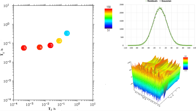
The Anahess T 1 –T 2 distribution from dried oat flakes either with the lowest BIC at 5 components (left) or at 4 components (right)
4 Conclusion
Provided that a dataset reflects a multi-exponential decay or recovery in one or two dimensions, the discrete Anahess processing tool returns a set of residual data or noise from the fit than can be regarded as symmetric and Gaussian. New raw datasets can then be produced from random permutations of the residuals, and they can be reprocessed using Anahess to find expectation values and standard deviations for T 1 , T 2 and the intensity. Also, the results from the fitted datasets can be used to find the likelihood of fitting to a certain number of components with the lowest BIC number.
Data Availability
No datasets were generated or analysed during the current study.
S.W. Provencher, CONTIN: A general purpose constrained regularization program for inverting noisy linear algebraic and integral equations. Comput. Phys. Commun. 27 (3), 229–242 (1982)
Article ADS Google Scholar
S.W. Provencher, A constrained regularization method for inverting data represented by linear algebraic or integral equations. Comput. Phys. Commun. 27 (3), 213–227 (1982)
S.W. Provencher, Inverse problems in polymer characterization: Direct analysis of polydispersity with photon correlation spectroscopy. Die Makromolekulare Chemie 180 (1), 201–209 (1979)
Article Google Scholar
L. Venkataramanan, Y. Song, M.D. Hürlimann, Solving Fredholm integrals of the first kind with tensor product structure in 2 and 2.5 dimensions. IEEE Trans. Signal Process. 50 , 1017–1026 (2002)
Article ADS MathSciNet Google Scholar
Y.Q. Song, L. Venkataramanan, M.D. Hürlimann, M. Flaum, P. Frulla, C. Straley, T1–T2 correlation spectra obtained using a fast twodimensional laplace inversion. J. Magn. Reson. 154 (2), 261–268 (2002)
G.C. Borgia, R.J.S. Brown, P. Fantazzini, Uniform-penalty inversion of multiexponential decay data. J. Magn. Reson. 132 , 65–77 (1998)
G.C. Borgia, R.J.S. Brown, P. Fantazzini, Uniform-penalty inversion of multiexponential decay data II. J. Magn. Reson. 147 , 273–285 (2000)
Teal P.D and Eccles C. Adaptive truncation of matrix decompositions and efficient estimation of NMR relaxation distributions. Inverse Problems 31 (4), (2015)
V. Villiam Bortolotti, L. Brizi, A. Nagmutdinova, F. Zama, and G. Landi, MUPen2DTool: A new Matlab Tool for 2D Nuclear Magnetic Resonance relaxation data inversion. SoftwareX 20 , 1–11 (2022)
Google Scholar
G.H. Sørland, H.W.A. AnthonsenUkkelberg, K. Zick, A robust method for analysing one and two-dimensional dynamic NMR data. Appl. Magn. Reson. 53 , 1345–1359 (2022)
Å. Ukkelberg, G.H. Sørland, E.W. Hansen, H.C. Widerøe, Anahess, a new second order sum of exponential fits, compared to the Tikhonov regularization approach, with NMR applications. Int. J. Recent Res. Appl. Stud. IJRRAS. 2 (3), 195–210 (2010)
P. Babak, S. Kryuchkov, A. Kantzas, Parsimony and goodness-of-fit in multi-dimensional NMR inversion. J. Magn. Reson. 274 , 46–56 (2017)
Yarman C.E, Monzón L., Reynolds M., Heaton N.: A new inversion method for NMR signal processing. In: 5th IEEE International Workshop on Computational Advances in Multi-Sensor Adaptive Processing (CAMSAP). 260–263 (2013)
S. Gideon, Estimating the Dimension of a Model. Ann. Stat. 6 (2), 461–464 (1978)
MathSciNet Google Scholar
G.H. Sørland, in Dynamic Pulsed-Field_Gradient NMR , (Springer Verlag, Berlin Heidelberg, 2014), pp. 139–145
A. O’Hagan, T. Leonard, Bayes estimation subject to uncertainty about parameter constraints". Biometrika 63 (1), 201–203 (1976)
Article MathSciNet Google Scholar
www.admagres.com . Accessed 1 Sept 2024 (2024)
www.antek.no . Accessed 1 Sept 2024 (2024)
G.H. Sørland, P.M. Larsen, F. Lundby, H.W. Anthonsen, B.J. Foss, On the Use of Low-Field NMR Methods for the Determination Of Total Lipid Content in Marine Products, in Magnetic Resonance in Food Science The Multivariate Challenge . (The Royal Society of Chemistry, 2005), pp.20–27
Chapter Google Scholar
Nordic-Baltic Committee on Food Analysis, "Fat determination in fish, fish feed and fish meal by low field nuclear magnetic resonance (LF-NMR)", NMKL 199, 2014
Download references
Open access funding provided by NTNU Norwegian University of Science and Technology (incl St. Olavs Hospital - Trondheim University Hospital).
Author information
Authors and affiliations.
Ugelstad Laboratory, Norwegian University of Science and Technology (NTNU), Trondheim, Norway
Geir Humborstad Sørland & Sebastien Simon
Anvendt Teknologi AS, Trondheim, Norway
Geir Humborstad Sørland & Henrik Walbye Anthonsen
You can also search for this author in PubMed Google Scholar
Contributions
All authors contributed to the study conception and design. Material preparation, data collection and analysis were performed by Geir Humborstad Sørland, Henrik Walbye Anthonsen, and Sebastien Simon. The first draft of the manuscript was written by Geir Humborstad Sørland and all authors commented on previous versions of the manuscript. All authors read and approved the final manuscript.
Corresponding author
Correspondence to Geir Humborstad Sørland .
Ethics declarations
Conflict of interest.
The authors declare no competing interests.
Additional information
Publisher's note.
Springer Nature remains neutral with regard to jurisdictional claims in published maps and institutional affiliations.
Rights and permissions
Open Access This article is licensed under a Creative Commons Attribution 4.0 International License, which permits use, sharing, adaptation, distribution and reproduction in any medium or format, as long as you give appropriate credit to the original author(s) and the source, provide a link to the Creative Commons licence, and indicate if changes were made. The images or other third party material in this article are included in the article's Creative Commons licence, unless indicated otherwise in a credit line to the material. If material is not included in the article's Creative Commons licence and your intended use is not permitted by statutory regulation or exceeds the permitted use, you will need to obtain permission directly from the copyright holder. To view a copy of this licence, visit http://creativecommons.org/licenses/by/4.0/ .
Reprints and permissions
About this article
Sørland, G.H., Anthonsen, H.W. & Simon, S. A Method for Analyzing Data from 1- and 2-Dimensional Relaxation and Diffusion NMR Experiments by Determination of their Expectation Values and Standard Deviations. Appl Magn Reson (2024). https://doi.org/10.1007/s00723-024-01718-z
Download citation
Received : 09 August 2024
Revised : 15 September 2024
Accepted : 16 September 2024
Published : 20 September 2024
DOI : https://doi.org/10.1007/s00723-024-01718-z
Share this article
Anyone you share the following link with will be able to read this content:
Sorry, a shareable link is not currently available for this article.
Provided by the Springer Nature SharedIt content-sharing initiative
- Find a journal
- Publish with us
- Track your research

IMAGES
VIDEO
COMMENTS
Resonance is a very important phenomenon in the study of vibrations, it occurs when an object is made to vibrate at its natural frequency by a nearby object ...
I show how our resonance tubes become louder or softer depending on the water level within. The difference between two resonant lengths is equal to one half...
Figure 16.25 shows an experiment you can try at home. Take a bowl of milk and place it on a common box fan. Vibrations from the fan will produce circular standing waves in the milk. ... The resonance produced on a string instrument can be modeled in a physics lab using the apparatus shown in Figure 16.28. Figure 16.28 A lab setup for creating ...
Resonance. The goal of Unit 11 of The Physics Classroom Tutorial is to develop an understanding of the nature, properties, behavior, and mathematics of sound and to apply this understanding to the analysis of music and musical instruments. Thus far in this unit, applications of sound wave principles have been made towards a discussion of beats ...
Super power? No, it's super science! Learn how to use the power of resonance to move objects with sound! Watch this video & tell us how your experiment went ...
The resonance produced on a string instrument can be modeled in a physics lab using the apparatus shown in Figure \(\PageIndex{4}\). ... Conducting this experiment in the lab would result in a decrease in amplitude as the frequency increases.) The next two modes, or the third and fourth harmonics, have wavelengths of \(\lambda_{3} ...
The relationship between these quantities is: v = fλ where. v = velocity of sound propagation. f = frequency. λ = wavelength. In this experiment the velocity of sound in air is to be found by using tuning forks of known frequency. The wavelength of the sound will be determined by making use of the resonance of an air column.
Resonance Experiment This three-minute YouTube video features David Goodstein from the Mechanical Universe series explaining and demonstrating the method of breaking a wine glass (or beaker) using resonance. The video segment includes excerpts from the usual 30-minute Lesson.
Demos: 4B-11 Resonance in Boxes with Tuning Forks. Two identical wooden boxes, open at one end, have identical tuning forks attached at the center of the top of the box. When the tuning fork is struck, the sound is amplified by the resonance occurring in the box. When the one box is placed such that its opening is a few centimeters from the ...
This laboratory experiment deals with acoustic resonance. For a more detailed treatment of waves and their basic properties, please see our standing wave experiment. Theory of Acoustic Resonance. Sound waves are longitudinal waves which require a medium, such as air or water, in which to travel. Sound does not travel in a vacuum.
Describe resonance and beats; Define fundamental frequency and harmonic series; Contrast an open-pipe and closed-pipe resonator; ... For this activity, switch to the Sound tab. Turn on the Sound option, and experiment with changing the frequency and amplitude, and adding in a second speaker and a barrier. According to the graph, what happens to ...
Resonance underlies aspects of the world as diverse as music, nuclear fusion in dying stars, and even the very existence of subatomic particles. Here's how the same effect manifests in such varied settings, from everyday life down to the smallest scales. ... Experiments indicated that light, which had been thought of as an electromagnetic ...
For advanced undergraduate students: Observe resonance in a collection of driven, damped harmonic oscillators. Vary the driving frequency and amplitude, the damping constant, and the mass and spring constant of each resonator. Notice the long-lived transients when damping is small, and observe the phase change for resonators above and below resonance.
The fundamental has λ = 4L, and frequency is related to wavelength and the speed of sound as given by: vw = fλ. Solving for f in this equation gives. f = vw λ = vw 4L, where vw is the speed of sound in air. Similarly, the first overtone has λ ′ = 4L / 3 (see Figure 17.5.9), so that. f ′ = 3vw 4L = 3f.
Strategy. The resonant frequency for a RLC circuit is calculated from Equation 15.6.5, which comes from a balance between the reactances of the capacitor and the inductor. Since the circuit is at resonance, the impedance is equal to the resistor. Then, the peak current is calculated by the voltage divided by the resistance.
1. Measure the length of the resonance tube. Record the length on the datasheet. 2. With the speaker placed a few centimeters from one end of the tube, as shown in Fig. 1 Connect the speaker to the "Low Ω" outputs of the frequency generator, as shown in Fig. 2 and connect the "High Ω" outputs to "CH1" of the oscilloscope.
Resonance is witnessed in objects in equilibrium with acting forces and could keep vibrating for a long time under perfect conditions. To find the resonant frequency of a single continuous wave, we use the formula, \ (\begin {array} {l}v = λf\end {array} \) Where v is the wave velocity and λ is the distance of the wavelength.
There are plenty of simple demonstrations of the resonance phenomena of mechanical systems that can be set up using readily available items. 1-5 Here I present a very simple approach to demonstrate the phenomena using just two spring scales. The experiment presented here performs a "frequency sweep" and is also very entertaining to watch.
Resonance Tube Equipment Capstone, complete resonance tube (tube, piston assembly, speaker stand, piston ... In another experiment, a pulse of sound will be sent down the tube and the re ected pulse examined. The nature of the re ected pulse will depend on whether the end of the tube is open or closed. Also, it will be possible to make a direct ...
Sign up for a free trial of The Great Courses Plus here: http://ow.ly/Dhlu30acnTCI use a flame tube called a Rubens Tube to explain resonance. Watch dancing ...
During an experiment of mechanics or physics, it is best to use scientific diagrams. For these subjects, experiments largely depend on the process. A correct setup and procedure can give an accurate result. Therefore, to learn about the tuning fork resonance experiment, the students should use scientific diagrams. However, it may be challenging ...
For the pulse experiments it is necessary to move the mike into the tube to the 10 cm mark. To do this, loosen the thumbscrew holding the mike. Move the speaker stand out a few cm from the resonance tube and then push the mike by its cord into the resonance tube. Then place the speaker stand back rmly against the resonance tube.
This is a better description of the electronic structure of ozone than either of the resonance structures in Figure \(\PageIndex{1}\). Many polyatomic ions also display resonance. In some cases, the true structure may be an average of three valid resonance structures, as in the case of the nitrate ion, \(\ce{NO_3^-}\) in Figure \(\PageIndex{3}\).
A diagram of the neutron-resonance transmission measurement setup using a laser driven neutron source (LDNS): (a) overview of the experimental setup with the neutron flight path; the sample used ...
When analyzing relaxation and/or diffusion data acquired with various Nuclear Magnetic Resonance (NMR) techniques followed by the use of the Inverse Laplace transform (ILT) [1,2,3,4,5,6,7,8,9], the smoothing of the data is essential to produce distributions of relaxation times and/or diffusion coefficients.Even though there has been some work in non-uniform smoothing, the most common way to ...
WARNING: Please lower your volume. The audio in this clip could cause hearing damage.Add me on Facebook - (click LIKE on Facebook to add me)http://www.facebo...